
")
Lisa Johnston, PhD, Product Expert at Volpara Health, explains the use of AI for mammography performance and quality enhancement
Within medical imaging, artificial intelligence (AI) is often seen as only a tool for detection and triage. However, an area where AI is already making a significant impact – for breast imaging centres and patients alike – is the assessment of mammography performance and image quality.
The mammogram is widely accepted as the screening exam that saves the most lives. (1) But mammography’s sensitivity depends on the quality of images taken, and image quality is impacted by differences in breast shape, overall body habitus, technologist skill, and patient tolerance for the procedure. These differences all directly impact patient positioning, reducing the mammogram’s effectiveness as a tool for the early detection of breast cancer. When image positioning quality fails to meet positioning criteria, mammography’s sensitivity drops from 84% to 66.3%. (2,3) Further, positioning issues – such as failing to include the entire breast on the mammographic image – can cause up to 80.6% of otherwise avoidable technical repeats/recalls. (4)
Given the importance of proper patient positioning, what measures are in place to ensure that high-quality mammogram images are obtained?
The challenge of achieving high-quality mammograms
The U.S. Federal Drug Administration (FDA) established national quality standards through the Mammography Quality Standards Act (MQSA) in the United States. Individual states also maintain their own quality standards. The standards include proper positioning, x-ray dose, and breast compression but, unfortunately, do not provide specific guidance on how to achieve the requisite high quality in these areas, nor how to sustain or improve performance.
At least two issues further complicate the situation: first, the subjective nature of conventional, visual assessment of positioning, due in part to both technologists’ inter-observer variability (1) and radiologists’ intra- observer variability; (2) and second, the lack of consensus on the evaluation criteria for good positioning. (3) It’s not surprising, then, that meeting the standards can prove challenging for some breast imaging centres, and even lead to operational consequences. According to the FDA, “[p]oor positioning has been found to be the cause of most clinical image deficiencies and most failures of accreditation.” (4)
To drive improvement in mammographic image quality, the FDA established the Enhancing Quality Using the Inspection Program (EQUIP) initiative. EQUIP inspections and American College of Radiology (ACR) accreditation both require imaging centres to provide evidence that their images meet MQSA standards.
Traditionally, collecting technologists’ best work has relied on visual assessment, a very time-consuming and subjective process that, in addition to being inconsistent between managers and not measurable, made sampling the only practical approach to monitoring quality.
This is the sweet spot where AI for mammography can do what humans cannot.
AI delivers consistent, objective quality assessment
AI excels in assessing large datasets that are too cumbersome for effective human processing. Volpara® AnalyticsTM software has so far analysed over 75 million images. Analytics is powered by the Volpara® TruPGMITM algorithm to automatically evaluate the image quality of every mammogram. TruPGMI was developed as an automated alternative to the PGMI Image Evaluation System, a visual assessment method published by the UK National Health System Breast Screening Programme. (5,6) Though they differ significantly in approach, both methods evaluate positioning and other features and use those features to classify mammographic studies as Perfect, Good, Moderate, or Inadequate (P, G, M, or I).
By assessing mammographic positioning against a set of precise metrics, TruPGMI provides an objective assessment of every image taken at a breast imaging facility. Evaluating every image provides a more accurate gauge of image quality than is possible from a sampling of images that may not represent the technologists’ best work or the patient population being screened.
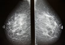
The circles on the image above indicate the areas that Volpara TruPGMI uses to assess the quality of each mammogram.
Such an AI approach to monitoring image quality yields considerable additional benefits. In this case, TruPGMI tracks image quality scores over time, providing analytics for each exam, technologist, mammography unit, and clinic, for both standard screening views. By understanding the positioning issues that affect image quality, breast imaging staff can identify key focus areas of improvement for individuals, teams, and sites. Managers and lead technologists can showcase high-quality work to improve morale. Technologists receive objective feedback that is consistent, fair, and measurable. Finally, managers spend significantly less time selecting images and preparing reports for ACR accreditation and EQUIP inspections. They can also highlight technologists’ excellent work, contributing to a workplace culture of quality.
Mayfair Diagnostics, a Calgary-based multi-modality imaging practice that takes over 50,000 mammograms annually, saw a 60% reduction in time searching for cases within six months of using Volpara Analytics – in addition to a 37% reduction in Inadequate images. (7)
The future of AI for mammography quality
AI’s role in mammographic quality assurance is the subject of numerous studies. Recently, Volpara Analytics was used to quantify mammography image quality improvement in the largest image quality evaluation to date.
With a dataset of over 48,000 exams conducted by 42 technologists over nine clinics, the study found that, in the 12 months following the installation of Analytics, there were significant improvements in breast positioning and compression quality – 6% and 8%, respectively. This led to an associated reduction in technical repeats and recalls of 78%. (8)
As the datasets grow larger and the studies more numerous, we expect to see more such success stories. But the real beneficiaries of the adoption of AI-powered mammography analytics are the patients. They will have the best chance of receiving a high-quality mammogram that includes all their breast tissue – and a much smaller likelihood of being called back because of low-quality images. Women should be confident in their mammography provider’s quality and their exam’s ability to afford them the best chance of early detection.
References 1-8 available upon request.

More About Stakeholder
-
 Volpara Health Limited – Saving more families from breast cancer
Volpara Health Limited provides clinically validated, AI-powered software for personalised screening and early detection of breast cancer.
Contributor Profile

Contributor Profile

Editor's Recommended Articles
-
-

-
 Must Read >> AI for breast imaging


 tools in genetics")


