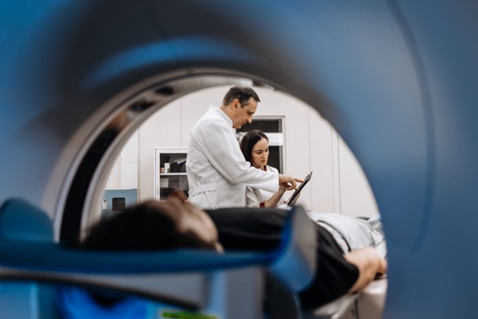
UKRI-funded researchers are using AI and Xenon gas imaging to make MRI scans faster, cheaper, and more accurate, improving lung disease diagnosis across the NHS
Artificial intelligence is transforming medical imaging, and UKRI-funded researchers are at the forefront of AI MRI scans that enhance the diagnosis of lung diseases. By combining AI algorithms with Xenon gas imaging, these advanced MRI techniques enable faster, more accurate, and more affordable scans. This innovation has the potential to enhance patient care across the NHS, helping doctors detect lung conditions earlier and deliver life-saving treatments more efficiently.
The technology was officially unveiled on September 8, 2025, at a ceremony held at the University of Sheffield’s MRI unit at the Royal Hallamshire Hospital.
Training AI MRI scanners
MRI scanners are widely used in all hospitals to create detailed images of the body and diagnose disease. However, these machines can be bulky and expensive, especially when large magnetic fields are used for advanced techniques.
Researchers from the POLARIS group and the University of Sheffield’s Insigneo Institute combined pioneering magnetic resonance imaging (MRI) lung-scanning technology with new MRI and artificial intelligence (AI) technology developed by GE HealthCare.
GE Healthcare trained AI software on images generated by MRI scanners, and the technology is used to reconstruct images from a low-field scanner with better quality than conventional algorithms, effectively achieving the quality of a high-field scan. This technique is more cost-effective, and more mobile MRI scans could be made available on the NHS, allowing more people to be seen more quickly, cutting waiting lists, and saving lives.
Pioneering Xenon gas in lung disease diagnosis
MRI scans successfully diagnose diseases in soft tissue; however, diseases of the lungs are more complex to diagnose because a significant portion of the lungs is composed of open space.
Now, researchers at the University of Sheffield have pioneered the use of imaging inhaled xenon gas in the lung space to generate high-quality gas MR images. The xenon gas is ‘hyperpolarised’ using Sheffield’s laser polarisation technology and can be used very effectively at low magnetic fields, and is being further developed as part of the project.
The combination of lung scanning with AI MRI technology is a world first and could improve the diagnostic technology for lung diseases.
Professor Jim Wild, Project Lead and Director of the Insigneo Institute, said: “Building, installing, and running MRI scanners gets more and more expensive the higher the magnetic field, and limits the accessibility and clinical reach of the technology. With advancements in engineering research, low-field MRI is experiencing a real comeback.
Combining our xenon MRI technology with cost-effective low-field AI MRI technology could mean a lot of people with respiratory disease get better and quicker access to a diagnosis and earlier treatment.”
Dr Jan Wolber, Global Product Leader Digital at GE HealthCare, said: “One of the significant benefits of AI technology is the cost-effectiveness for the NHS. It could mean you will be able to do more with the same money.
If these were installed across hospitals, they should help reduce waiting lists. Patients could be seen more quickly after referral from a GP. Earlier diagnosis in oncology typically leads to better outcomes, potentially saving or prolonging lives.
We often push for more complex and feature-rich systems. Health services are under cost pressure, so sometimes we need to revisit whether we can achieve the same outcome for less money. Advances in AI allow that, and this is why it is so impactful.”


