Chima V. Maduka, DVM, MS, PhD, and Christopher H. Contag, PhD, provide insights on tuning immunometabolism to resolve inflammation and promote repair at the bone–biomaterial interface
Host immune responses ultimately dictate the success or failure of biopolymer implants used in tissue repair. After a bone injury, the ideal response to an implanted biomaterial is brief, coordinated inflammation followed by resolution, vascularization, and remodeling. Too often, however, the response becomes chronic: neutrophils persist, monocyte-derived cells skew inflammatory responses, dendritic cells remain activated, and the foreign-body reaction hardens into a barrier against integration. The central question is no longer whether the immune system matters – it’s how to steer it. A compelling answer is emerging from a recent series of papers from our laboratories, revealing that immune cells can be reprogrammed by targeting their metabolism at the biomaterial interface.
From material chemistry to immune metabolism
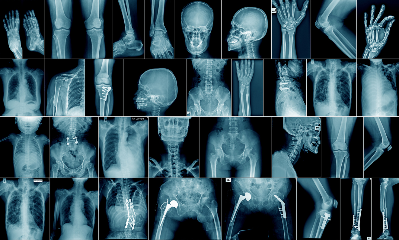
Classically, adverse reactions to bone biomaterials (e.g., polylactide–hydroxyapatite composites) were pinned on surface roughness, particle size, crystallinity, and pH. Those factors still matter, but we have revealed a deeper lever: the metabolic state of immune cells encountering degrading polymers. We show that polylactide (PLA) degradation products shift macrophages and other myeloid cells toward glycolysis and associated inflammatory programs; conversely, interrupting glycolytic flux can tilt the microenvironment toward repair. We demonstrated that PLA degradation products activate immune cells via metabolic reprogramming, reframing ‘biocompatibility’ measures to include immunometabolic endpoints and immune signaling. (1)
PLA–hydroxyapatite (HA) composites are widely used for bone, cartilage, and dental repair, and we showed that metabolic inhibitors embedded in amorphous PLA (aPLA) + HA composites tune the glycolytic flux in situ and guide the regenerative response. The outcome was a coordinated shift in the peri-implant immune landscape, characterized by reduced neutrophils and pro-inflammatory monocytes/ macrophages (CD86+CD206-), increased CD206+ pro-regenerative cells, and higher arginase-1 expression among dendritic cells – changes that are expected to favor angiogenesis and osseointegration. In short, controlling local immunometabolism created a pro-regenerative niche around a clinically relevant bone-repair composite. (2)
Trafficking is also metabolic
Beyond macrophage polarization at the tissue-implant interface, metabolic cues regulate which cells are recruited into the tissue site. Using dual-reporter mice (CCR2-RFP and CX3CR1-GFP), intravital imaging, and metabolically modulated PLA implants, we linked the local metabolic state to the chemokine signals that recruit cells, including CCR2/CX3CR1-dependent trafficking of neutrophils and monocytes into the biomaterial microenvironment.
This metabolic gating set the stage for the downstream assembly of pro-inflammatory, transitional, and anti-inflammatory/pro-regenerative myeloid populations. Notably, glycolytic inhibition with 2-deoxy-D-glucose (2DG) embedded within amorphous PLA (aPLA) reduced glucose utilization, as demonstrated by fluorodeoxyglucose (FDG) radiotracer uptake and positron emission tomography (PET) imaging, and redirected immune responses toward resolution. In crystalline PLA (cPLA), type-2 helper T cells and IL-4–producing γδ T cells collaborated with arginase-1+ myeloid cells to establish a pro-healing environment – highlighting that material structure and drug release kinetics shape the immunometabolic ‘grammar’. (3)
Together, these studies crystallize a unifying principle: the biomaterial doesn’t just present a surface; it presents a metabolic context, and lactate monomers and oligomers act as signaling molecules at the tissue-implant interface, much like in cancer, driving a metabolic shift toward glycolysis. This context can be chemically modified. The metabolic context dictates the recruitment, activation state, and crosstalk among innate and adaptive immune cells, which ultimately determine whether bone integrates or scars.
Why immunometabolism matters in bone repair
Bone regeneration demands a precisely timed handoff: early inflammatory signals (often glycolysis-biased) must yield to oxidative, pro-resolving programs that support angiogenesis, osteoprogenitor recruitment, and matrix deposition. Chronic glycolytic drive at the interface locks macrophages and dendritic cells into danger signaling modes, keeps neutrophils present, and sustains cytokine circuits that impair osteogenesis. By locally dialing down glycolysis with inhibitors targeting hexokinase (using the drug 2-deoxyglucose), transaminases (with the drug aminooxyacetic acid),
or at other metabolic nodes, we were able to rebalance that immune timing and diminish the immune response. Notably, by delivering the material (rather than systemic immunosuppression) concentrates the effect where it is needed while minimizing off-target risk. (2)
This data also bridges soft-tissue and orthopedic contexts. Prior observations that polyethylene wear particles drive glycolytic reprogramming of innate immune cells in periprosthetic inflammation suggest a common immunometabolic thread across arthroplasty failure and degradable polymer implants. (4,5)
Design principles for next-gen bone biomaterials
Our findings suggest an actionable playbook for designing bone-repair constructs:
- Build in metabolic feedback. Incorporate metabolic modulators at low, sustained doses to adjust glycolytic/oxidative balance as the material degrades – even modest release can reshape immune composition and function. (2)
- Match inhibitor kinetics to polymer physics. aPLA and cPLA release drugs at different rates; the latter’s slow kinetics revealed distinct immune choreography (e.g., Th2/γδ T-cell IL-4 contributions). This argues for tuning crystallinity, geometry, and porosity alongside drug loading. (3)
- Engineer early trafficking, not only polarization. CCR2/CX3CR1-linked recruitment can be nudged by metabolic state. Guiding which cells arrive when may be as critical as pushing M1→M2 macophage transitions after arrival. (3)
- Use metabolic imaging as a go/ no-go gate. The observation that FDG-PET around 3D-printed PLA femoral repairs drops with glycolytic inhibition suggests a noninvasive biomarker to iterate formulations preclinically and, potentially, clinically. (3)
- Consider composites holistically. HA can promote osteogenesis but may amplify neutrophil recruitment, depending on the formulation; immunometabolic tuning can offset these liabilities and unlock HA’s benefits. (2)
Translational path and clinical promise
For segmental defects and challenging nonunions, surgeons increasingly turn to patient-specific, 3D-printed scaffolds and PLA-based fixation. Our work suggests that the scaffolds serve to bear the load and can also guide osteoconduction by shaping local immunity toward vascularized, osteogenic healing. Because the targets (e.g., hexokinase) and readouts (FDG-PET) are already familiar in oncology and inflammation imaging, the regulatory path may be shorter than for wholly novel biologics.
Key translational steps
Dose-finding and safety studies for embedded metabolic modulators must carefully consider local concentration, release duration, and clearance. To validate translational relevance, large-animal bone-defect models that recapitulate human-scale mechanics and immunology are needed to demonstrate that immunometabolic steering promotes osseointegration, trabecular bridging, and restoration of mechanical strength. Furthermore, patient stratification will be critical, as systemic metabolic states such as diabetes and obesity, as well as prior implant exposures, may condition baseline immune metabolism; thus, local modulation strategies could be tailored for personalized therapeutic benefit.
Guiding the field
The case for immunometabolic control is strong, but several guiding questions remain. It is not yet clear which metabolic node offers the most effective intervention; while hexokinase inhibition has shown promise, alternative strategies such as modulating lactate handling, glutamine metabolism, or tricarboxylic cycle (TCA) shunts may provide finer control or fewer trade-offs depending on defect size, vascularity, and how adaptive immune cells use these signals. Results from crystalline PLA models implicating Th2 and γδ T-cell IL-4 suggest that myeloid tuning can propagate to T-cell programs critical for bone repair. Mapping this cascade, in both time and space, could reveal synergies with cytokine-releasing coatings. It is likely that an early, strong blockade of glycolysis, aimed at restraining neutrophils, followed by a gradual taper that permits remodeling, would be beneficial. Smart materials capable of sensing inflammation and releasing inhibitors on demand will help guide this process. The osteoconductive properties of hydroxyapatite, combined with immunometabolic steering, may outperform either strategy alone.
The addition of BMP-mimetic peptides or pro-angiogenic factors, aligned with the immune curve, could further accelerate bone union.
From foreign-body reaction to cooperative remodeling
We have repositioned the implant interface as a metabolically programmable ecosystem. By embedding modest doses of metabolic modulators in clinically relevant PLA/HA composites and showing effects on cell trafficking, phenotype, and imaging-visible inflammation, it is possible to convert a persistent liability – chronic inflammation – into a design variable. For bone repair, where blood supply, immune choreography, and mechanical environment must align, immunometabolic targeting offers a precise, local, and material-native lever to shift outcomes from fibrotic encapsulation to vascularized osseointegration and robust remodeling. The message for translational teams is clear: treat immunometabolism as a first-class design constraint for bone biomaterials. Choose polymer chemistry and crystallinity with drug-release kinetics in mind, bias early CCR2/CX3CR1-dependent trafficking toward constructive players, and verify success with functional metabolic imaging. When materials learn to “speak metabolism,” the bone learns to heal.
References
- Maduka, C. V., Alhaj, M., Ural, E., Habeeb, O. M., Kuhnert, M. M., Smith, K., Makela, A. V., Pope, H., Chen, S., Hix, J. M., Mallett, C. L., Chung, S. J., Hakun, M., Tundo, A., Zinn, K. R., Hankenson, K. D., Goodman, S. B., Narayan, R. & Contag, C. H. Polylactide Degradation Activates Immune Cells by Metabolic Reprogramming. Adv Sci (Weinh) 10, e2304632, doi:https://doi.org/10.1002/advs.202304632 (2023).
- Maduka, C. V., Makela, A. V., Tundo, A., Ural, E., Stivers, K. B., Kuhnert, M. M., Alhaj, M., Hoque Apu, E., Ashammakhi, N., Hankenson, K. D., Narayan, R., Elisseeff, J. H., & Contag, C. H. Regulating the proinflammatory response to composite biomaterials by targeting immunometabolism. Bioact Mater 40, 64-73, doi:https://doi.org/10.1016/j.bioactmat.2024.05.046 (2024).
- Maduka, C. V., Schmitter-Sanchez, A. D., Makela, A. V., Ural, E., Stivers, K. B., Pope, H., Kuhnert, M. M., Habeeb, O. M., Tundo, A., Alhaj, M., Kiselev, A., Chen, S., Donneys, A., Winton, W. P., Stauff, J., Scott, P. J. H., Olive, A. J., Hankenson, K. D., Narayan, R., Park, S., Elisseeff, J. H. & Contag, C. H. Immunometabolic cues recompose and reprogram the microenvironment around implanted biomaterials. Nat Biomed Eng 8, 1308-1321, doi: https://doi.org/10.1038/s41551-024-01260-0 (2024).
- Teissier, V., Gao, Q., Shen, H., Li, J., Li, X., Huang, E. E., Kushioka, J., Toya, M., Tsubosaka, M., Hirata, H., Alizadeh, H. V., Maduka, C. V., Contag, C. H., Yang, Y. P., Zhang, N. & Goodman, S. B. Metabolic profile of mesenchymal stromal cells and macrophages in the presence of polyethylene particles in a 3D model. Stem Cell Res Ther 14, 99, doi:https://doi.org/10.1186/s13287-023-03260-4 (2023).
- Maduka, C. V., Habeeb, O. M., Kuhnert, M. M., Hakun, M., Goodman, S. B., & Contag, C. H. Glycolytic reprogramming underlies immune cell activation by polyethylene wear particles. Biomater Adv 152, 213495, doi: https://doi.org/10.1016/j.bioadv.2023.213495 (2023).