Researchers at King’s College London have developed an AI model that can detect strokes, brain tumours, and aneurysms from MRI scans, helping doctors make faster, more accurate diagnoses
Researchers at King’s College London have made a significant advance in medical imaging by developing an AI model that accurately detects strokes, brain tumours, and aneurysms in MRI scans. This groundbreaking technology aims to enhance the speed and accuracy of diagnoses, reduce radiologists’ workload, and ultimately improve patient outcomes by enabling faster identification of critical cases requiring immediate attention. The detailed findings of this study have been published in the reputable journal Radiology AI.
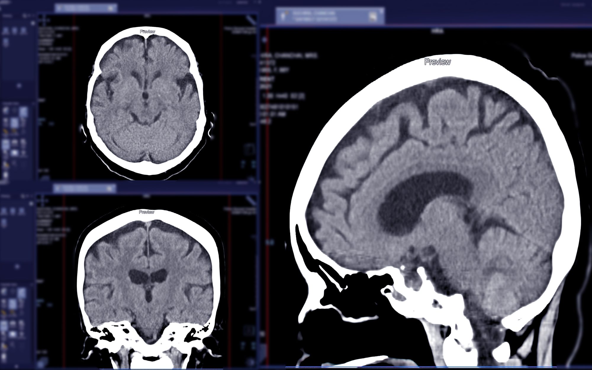
New AI tool to address increasing pressure in radiology
The newly developed AI tool is designed to address the increasing pressure on radiology departments by efficiently triaging scans and expediting reporting. To achieve this, the model underwent initial training to distinguish between ‘normal’ and ‘abnormal’ scans. Impressively, it demonstrated high accuracy comparable to that of expert radiologists, showcasing its potential as a reliable diagnostic assistant.
In a series of rigorous tests, the AI model was evaluated on its ability to identify specific neurological conditions, including strokes, multiple sclerosis, and brain tumours. This testing involved using MRI scans that were not included in the model’s training dataset. Remarkably, the AI successfully recognised these conditions with high precision, confirming its robustness in clinical settings.
A distinguishing feature of this AI model is its training methodology. Unlike many traditional AI models that rely on large, manually labeled datasets, the researchers employed a self-learning approach by utilising over 60,000 MRI scans. This innovative technique not only reduced reliance on extensive manual labeling but also increased the model’s ability to adapt and improve autonomously, thereby inspiring confidence in its diagnostic capabilities.
The researchers have also designed the AI system to serve as a valuable tool in both diagnostic reviews and educational settings. By effectively searching for and retrieving similar cases from a database of past examinations, the AI could facilitate more effective learning and decision-making for medical professionals. This capability has the potential to significantly streamline healthcare workflows. In practice, the AI model could be integrated into MRI scans to flag abnormalities in real time, assist radiologists by suggesting potential findings, identify potential discrepancies in reports, and provide access to comparable cases from previous studies. Such functionalities would not only accelerate the diagnostic process but also minimise reporting delays.
Detecting abnormalities on scans
Dr. Thomas Booth, who is a Reader in Neuroimaging at King’s College London and a Consultant Neuroradiologist at King’s College Hospital, explained the significance of this approach: “By training the system on scans and the language radiologists use to describe them, we can teach it to understand what abnormalities look like.”
Looking ahead, the researchers are eager to take the next step toward wider clinical application by conducting a randomised multicenter trial across hospitals in the UK. This trial will aim to evaluate how the AI’s ability to detect abnormalities improves diagnostic workflows in real-world practice. Dr. Booth expressed enthusiasm for the upcoming phase of research, stating, “The next step is to run a randomised multicentre trial across the UK to see how abnormality detection improves workflows in practice. We are pleased to say that this trial will start in hospitals in 2026.”


