Open Access Government produces compelling and informative news, publications, eBooks, and academic research articles for the public and private sector looking at health, diseases & conditions, workplace, research & innovation, digital transformation, government policy, environment, agriculture, energy, transport and more.
Home 2025
Archives
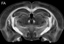
3D microscopic whole brain neurodegenerative MRI
This article by G. Allan Johnson, Ph.D., focuses on advanced MRI techniques for studying neurodegenerative diseases, exploring the challenges of screening therapies for conditions like Alzheimer’s and Parkinson’s, and highlighting the promising research conducted at Duke University.


