Dr Adèle Ehongo discusses peripapillary intrachoroidal cavitation (PICC), a masquerade of normal-tension glaucoma
When it looks like normal tension glaucoma but isn’t, simply consider unmasking it with Optical Coherence Tomography (OCT).
Glaucoma is the leading cause of irreversible blindness worldwide. (1) Normal-tension glaucoma is a subtype characterized by progressive optic nerve damage despite intraocular pressure remaining within the normal range. (1) This diagnosis is made after excluding other causes of glaucomatous-type visual field defects (VFDs). In this context, peripapillary intrachoroidal cavitation (PICC) should be excluded, which has recently gained renewed interest thanks to advances in OCT. (2,3,4)
Asymptomatic, but concerning
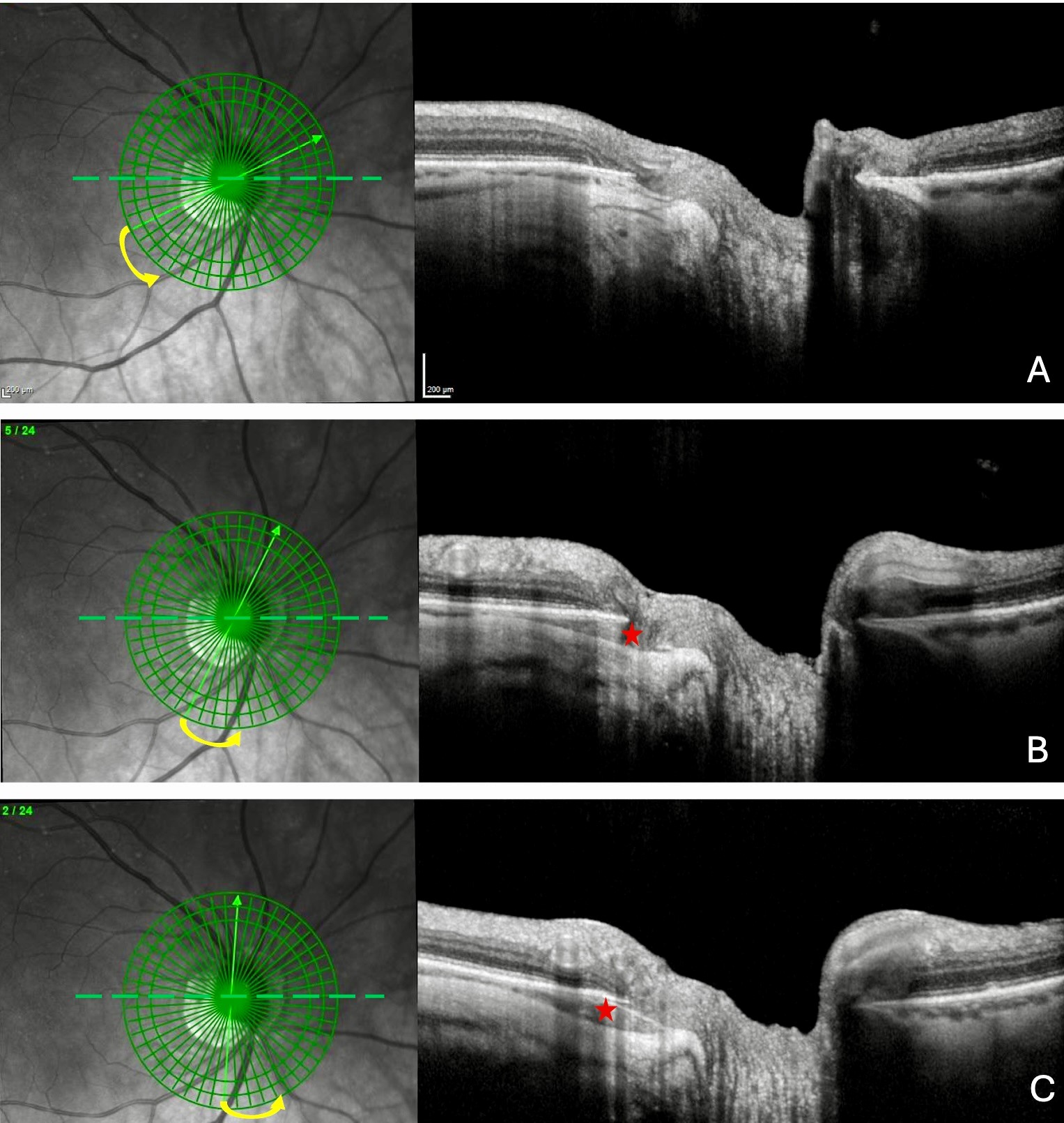
With a prevalence of up to 22% in highly myopic eyes, (5) PICC is a yellow-orange lesion located at the outer limit of the myopic conus (4,6) (Figure 1.A). Although PICC is usually asymptomatic, it may be concerning, suggesting a choroidal tumour. (6)
Invisible, and therefore confusing
However, the yellow-orange appearance is absent in up to 53% of PICCs detected on OCT. (7) Since PICCs are associated with VFDs similar to those of glaucoma in up to 73.3% of cases, (8) this can lead to diagnostic confusion and overtreatment. Although PICC-related VFDs rarely progress, glaucoma, as an irreversible cause of blindness, requires timely diagnosis and treatment, necessitating rapid distinction from other causes of VFDs.
Non-causal association
Despite the relationship between PICC and VFDs and although myopic eyes with PICC exhibit more VFDs than eyes without this condition, (4,9) mechanisms leading to the appearance of VFDs in eyes with PICCs remain to be established. Rare cases of documented full thickness defects (Figure 1.A and B) accompagnying certain PICCs explain some visual field defects. The PICC itself, as a suprachoroidal detachment, cannot explain these deficits.(3,4,9) However, PICCs and VFDs may reflect the impact of a promotor acting around the optic nerve, distorting structures and inducing the development of VFDs. (2-4,9) This promotor was suggested to be the forces exerted by optic nerve sheaths at their scleral attachments. (2-4,9,10)
It is rewarding to dig in easily, quickly, and unmask PICC
PICC diagnosis is simple, fast, and cost-effective, with a single click, thanks to ready-to-use radial OCT slices for glaucoma assessment (2-4) (Figure 2). By keeping the PICC in mind, the clinician avoids multiple diagnostic investigations dictated by the presence of the yellow-orange aspect. When the PICC is not visible, the clinician is also not misled about the diagnosis of glaucoma, which could even lead to unnecessary surgery.
Promising
The pathogenic mechanism of PICC, related to the traction of the optic nerve sheaths on their attachments, is relevant to other myopic complications (2-4,10), warranting further study.
References
- Jayaram H, Kolko M, Friedman DS, Gazzard G. Glaucoma: now and beyond. Lancet. 2023 Nov 11;402(10414):1788-1801. doi: https://doi.org/10.1016/S0140-6736(23)01289-8. Epub 2023 Sep 21. PMID: 37742700.
- Ehongo A. Doctoral thesis in Medical science (2024). Free University of Brussels. Optical Coherence Tomography analysis of Peripapillary intrachoroidal cavitation. Clinical implications for highly myopic eyes.
- Ehongo A, Bacq N, Kisma N, Dugauquier A, Alaoui Mhammedi Y, Coppens K, Bremer F, Leroy K. Analysis of Peripapillary Intrachoroidal Cavitation and Myopic Peripapillary Distortions in Polar Regions by Optical Coherence Tomography. Clin Ophthalmol. 2022 Aug 13;16:2617-2629. doi: https://doi.org/10.2147/OPTH.S376597. PMID: 35992567; PMCID: PMC9387167.
- Ehongo A, Bacq N. Peripapillary Intrachoroidal Cavitation. J Clin Med. 2023 Jul 16;12(14):4712. doi: https://doi.org/10.3390/jcm12144712. PMID: 37510829; PMCID: PMC10380777.
- Choudhury F, Meuer SM, Klein R, Wang D, Torres M, Jiang X, McKean-Cowdin R, Varma R; Chinese American Eye Study Group. Prevalence and Characteristics of Myopic Degeneration in an Adult Chinese American Population: The Chinese American Eye Study. Am J Ophthalmol. 2018 Mar;187:34-42. doi: https://doi.org/10.1016/j.ajo.2017.12.010. Epub 2017 Dec 27. PMID: 29288031; PMCID: PMC5837945.
- Freund KB, Ciardella AP, Yannuzzi LA, Pece A, Goldbaum M, Kokame GT, Orlock D. Peripapillary detachment in pathologic myopia. Arch Ophthalmol. 2003 Feb;121(2):197-204. doi: https://doi.org/10.1001/archopht.121.2.197. PMID: 12583785.
- Dai Y, Jonas JB, Ling Z, Wang X, Sun X. Unilateral peripapillary intrachoroidal cavitation and optic disk rotation. Retina. 2015 Apr;35(4):655-9. doi: https://doi.org/10.1097/IAE.0000000000000358. PMID: 25299968.
- Okuma S, Mizoue S, Ohashi Y. Visual field defects and changes in macular retinal ganglion cell complex thickness in eyes with intrachoroidal cavitation are similar to those in early glaucoma. Clin Ophthalmol. 2016 Jun 29;10:1217-22. doi: https://doi.org/10.2147/OPTH.S102130. PMID: 27418805; PMCID: PMC4935007.
- Ehongo A, Dugauquier A, Kisma N, De Maertelaer V, Nana Wandji B, Tchatchou Tomy W, Alaoui Mhammedi Y, Coppens K, Leroy K, Bremer F. Myopic (Peri)papillary Changes and Visual Field Defects. Clin Ophthalmol. 2023 Nov 1;17:3295-3306. doi: https://doi.org/10.2147/OPTH.S404167. PMID: 37933329; PMCID: PMC10625749.
- Ehongo A. Understanding Posterior Staphyloma in Pathologic Myopia: Current Overview, New Input, and Perspectives. Clin Ophthalmol. 2023 Dec 12;17:3825-3853. doi: https://doi.org/10.2147/OPTH.S405202. PMID: 38105912; PMCID: PMC10725704.