
Recent development in in situ electron microscopy is discussed by Thomas Willum Hansen, Senior Researcher at DTU CEN – Center For Electron Nanoscopy.
Contemporary society requires the continued need for optimized materials with a performance always surpassing the previous generation with at least an order of magnitude. In order to design, engineer and synthesize such materials, knowledge of the position of each atom in the structure is highly beneficial. In addition, such knowledge should be extracted with the materials as close as possible to its working state as discrepancies in atomic configuration can be highly environment dependent. Such investigations are known as in situ studies.
Recent development in in situ electron microscopy include heaters allowing for rapid heating to beyond 1000°C, gas cells that can maintain atmospheres higher than atmospheric pressure (while imaging), and cameras capable of frame rates exceeding 300 frames per second. Using such technologies in conjunction with state-of-the-art transmission electron microscopes (TEM) able to resolve distances smaller than 0.1 nm, researchers can pinpoint atoms in nanostructures with unprecedented resolution, even under extremely challenging conditions (Hansen and Wagner 2016). Perhaps even more interesting, changes in nanoscale systems are observed with an unprecedented spatial and temporal resolution. In combination with in situ imaging, spectroscopy in the form energy dispersive X-ray spectroscopy and electron energy-loss spectroscopy can be carried out ensuring the chemical information.
We have investigated the reactions occurring at the surfaces transition metals such as nickel, gold, copper and platinum. These elements are all highly relevant for various catalytic applications ranging from automotive exhaust abatement to growth of single graphene sheets. The aim of this investigation is to unravel the surface states leading to specific reaction pathways.
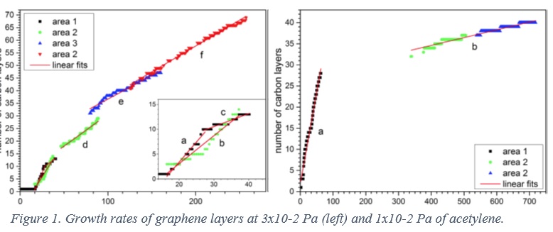
In the case of graphene growth, the target is to optimize the growth conditions towards high growth rates while maintain a low number if defects (Kling, Hansen et al. 2016). The main observations form this work shows that the growth rate varies as a function of how many layers of graphene has been grown. As the number of graphene layers increase, the growth rate of additional layers decreases, see Fig. 1. As the pressure of the carbon precursor is decreased, this effect is more evident. Such observations can guide the manufacturers of graphene to achieve a specific type of material with a specific number of graphene layers by tuning the growth conditions.
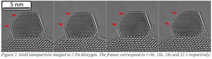
One of the holy grails within the study of catalytic materials is observing the site on the catalyst at which reactant species adsorb and react to form products. This site is known as the active site. In situ investigations of catalytic materials show that catalysts are active materials at all levels ranging from pellet scale (10-2 m) down to the atomic level (Hansen, Wagner et al. 2001). This means that the active site is not a stable configuration of surface atoms. This is exemplified in Fig. 2 where a temporally resolved image series of a gold nanoparticle (a material that can convert carbon monoxide and molecular oxygen to carbon dioxide) is imaged in the presence of 2 Pa molecular oxygen. The sites with less neighboring atoms appear to be less rigid and the atomic configuration changes from frame to frame, a phenomenon not observed in vacuum. This indicates that the oxygen is inducing changes on the surface at the atomic level. The net result of the atoms moving around on the surface of the nanoparticle can be mobility of the particle as a whole. Such motion can be detrimental to the lifetime of the catalyst (Hansen, Delariva et al. 2013).
In summary, using high-resolution in situ electron microscopies it is possible to gain an insight in the active state of nanomaterials. Mobile atoms can are resolved with high spatial and temporal resolution in environments resembling an operational scenario for the materials in question. Such observations could guide future development in fields such as catalysis and functionalized materials, and at the same time provide new knowledge of the interaction between gases and solids.
Hansen, T. W., A. T. Delariva, S. R. Challa and A. K. Datye (2013). “Sintering of Catalytic Nanoparticles: Particle Migration or Ostwald Ripening?” Accounts of Chemical Research 46(8): 1720-1730.
Hansen, T. W. and J. B. Wagner, Eds. (2016). Controlled Atmosphere Transmission Electron Microscopy, Springer.
Hansen, T. W., J. B. Wagner, P. L. Hansen, S. Dahl, H. Topsøe and C. J. H. Jacobsen (2001). “Atomic-resolution in situ transmission electron microscopy of a promoter of a heterogeneous catalyst.” Science 294(5546): 1508-1510.
Kling, J., T. W. Hansen and J. B. Wagner (2016). “Quantifying the growth of individual graphene layers by in situ environmental transmission electron microscopy.” Carbon 99: 261-266.
Senior researcher Thomas Willum Hansen
Center for Electron Nanoscopy
DTU Cen, Technical University of Denmark
Tel: +45 4525 6476
thomas.w.hansen@cen.dtu.dk
www.cen.dtu.dk
www.danchip.dtu.dk
www.twitter.com/dtu_danchip_cen


