Johns Hopkins researchers have grown a multi-region human brain organoid with rudimentary blood vessels and connected neural circuits that mimic fetal brain development
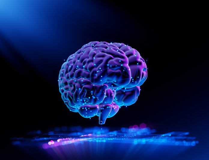
In a significant leap for neuroscience, researchers at Johns Hopkins have grown a miniature human brain in the lab that mimics key features of real brain development. This multi-region organoid not only forms functional neural connections but also glows as it transmits signals, offering a powerful new model for studying conditions like autism, schizophrenia, and Alzheimer’s disease.
“We’ve made the next generation of brain organoids,” said lead author Annie Kathuria, an assistant professor in JHU’s Department of Biomedical Engineering who studies brain development and neuropsychiatric disorders. “Most brain organoids that you see in papers are one brain region, like the cortex, the hindbrain or the midbrain. We’ve grown a rudimentary whole-brain organoid; we call it the multi-region brain organoid (MRBO).”
The findings are detailed in Advanced Science.
A world-first for scientists
The research marks one of the first times scientists have generated an organoid with tissues from each region of the brain connected and acting in concert.
To create a whole-brain organoid, Kathuria and her team first grew neural cells from the separate regions of the brain and rudimentary forms of blood vessels in separate lab dishes. The researchers then stuck the individual parts together with sticky proteins that act as a biological superglue and allowed the tissues to form connections. As the tissues began to grow together, they started producing electrical activity and responding as a network.
The multi-region mini brain organoid retained a broad range of types of neuronal cells, with characteristics resembling a brain in a 40-day-old human fetus. Some 80% of the range of the kinds of cells commonly seen at the early stages of human brain development was equally expressed in the laboratory-crafted miniaturised brains. Much smaller compared to a real brain, weighing in at 6 million to 7 million neurons compared with tens of billions in adult brains, these organoids provide a unique platform on which to study whole-brain development.
The researchers saw the creation of an early blood-brain barrier formation, which is the layer of cells surrounding the brain that controls which molecules can pass through.
“We need to study models with human cells if you want to understand neurodevelopmental disorders or neuropsychiatric disorders, but I can’t ask a person to let me take a peek at their brain just to study autism,” Kathuria said. “Whole-brain organoids let us watch disorders develop in real time, see if treatments work, and even tailor therapies to individual patients.”
New targets for drug screening
Whole-brain organoids could be a beacon of hope in drug development, potentially improving the success rate of clinical trials and saving money. This is vital as around 85% to 90% of drugs fail during Phase 1 clinical trials. For neuropsychiatric drugs, the failure rate is closer to 96%. The failure rate is high as scientists mostly use animal models during the early stages of drug development, which, unlike whole-brain organoids, do not closely resemble the natural development of the human brain.
“Diseases such as schizophrenia, autism, and Alzheimer’s affect the whole brain, not just one part of the brain. If you can understand what goes wrong early in development, we may be able to find new targets for drug screening,” Kathuria said. “We can test new drugs or treatments on the organoids and determine whether they’re having an impact on the organoids.”


