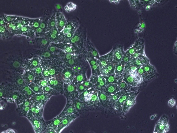
At the heart of almost everything we do in chemistry is analysis, measurements that show us what we have made, how pure it is, what is happening in a reaction, or what is present in a particular sample. The use of the word ‘see’ is not just a journalistic title because a very high proportion of analytical methods rely upon the absorption or emission of light and the sensitivity of some modern analytical technologies is sufficient to detect just a few molecules of a compound in a sample. If you’ve read any of my preceding Special Reports you’ll expect that heterocyclic compounds are at the heart of these methods and this is indeed the case. The variety and use of heterocyclic compounds as key components of analytical methods and devices is astounding. In our work searching for new treatments for drugs to treat prostate and pancreatic cancer we routinely use assays that involve the emission of light to determine whether our compounds have the basic requirements to evaluate further as drugs. These assays use a heterocyclic compound known as a luciferin, which emits light when it is oxidized; the light emission can be measured and its intensity tells you how much of the material you are analyzing is present. We can see more literally where our molecules go using fluorescence. Some of our antibacterial and antiparasitic drugs are fluorescent and in understanding how they work it’s very helpful to see whether they are taken up into cells and where they go in the cell. This helps to define the selectivity of compounds between bacterial cells, parasitic cells, and healthy mammalian cells, for example, so that we can get a safe drug. Such a case is shown in the figure where we can see a probe compound through its green fluorescence having migrated into the nuclei of the liver cells.
And it’s not just in the laboratory of the professional scientist that ‘seeing’ molecules is important. The commercial development of clinical analytical devices is proceeding apace and some of my colleagues at the University of Strathclyde are at the forefront of these developments. Professor Duncan Graham and Dr Karen Faulds lead a large team of researchers housed in the magnificent new Technology and Innovation Centre at the University of Strathclyde. Their group, known as the Centre for Molecular Nanometrology, is world leading in the science and application of a technique known as Surface Enhanced Raman Spectroscopy, or SERS for short. Using this technology they have developed many analytical methods with the potential for the development in clinically useful assays. It is possible to identify specific genes and specific pathogens, in some cases several at a time. These methods have been developed to be sufficiently robust for commercial application and their spin-out company, Renishaw Diagnostics, is now promoting a method and machine for the simultaneous analysis of twelve different fungal pathogens. Other applications being developed include the detection of viruses and the identification of bacteria that cause gastroenteritis. It’s remarkable what you can see using the right chemistry!


