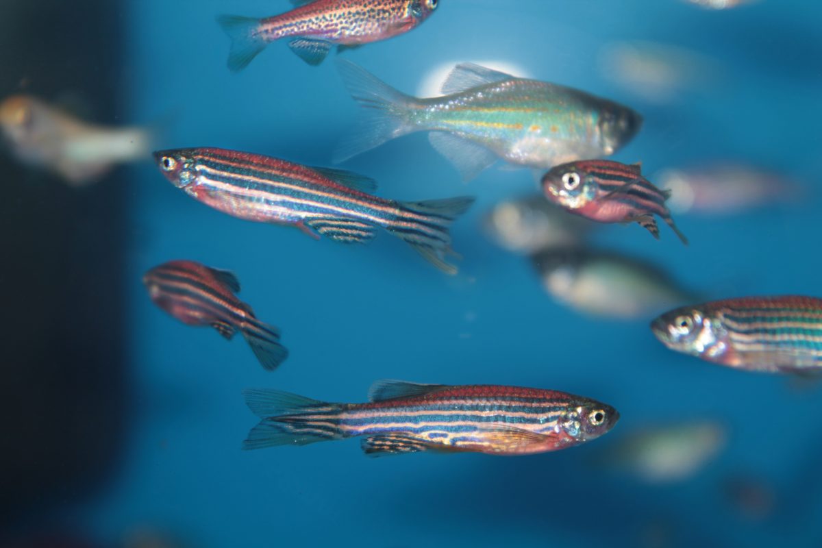
Christopher L Antos, Group Leader at the DFG-Center for Regenerative Therapies Dresden explains how Zebrafish research is a vital tool to help understand organ and appendage regeneration.
Injuries from trauma, ischemia and amputation can result in the permanent loss of an organ and limb function. The regeneration of many vital organs and appendages is a phenomenon that is limited or absent in humans, and this limitation can result in severe incapacitation or mortality. Such outcomes generate a medical need to find strategies to replace tissue loss. These strategies may come from understanding the cell biology and molecular mechanisms that are used by animals which have a significant capacity to regenerate organs and appendages, and there are vertebrates that have the ability to regenerate with such high fidelity that it is impossible to identify the original loss. While it remains unclear why some animals can regenerate specific structures and others cannot, what current research is finding is that many of the molecular mechanisms involved in regeneration are involved in embryonic development and these basic molecular mechanisms are conserved among the species. Thus, it should be possible to partially or completely restore the human endogenous ability to regenerate appendages and organs by providing the missing signals identified through studies of those animals that regenerate.
The processes governing regeneration are very complex. After injury, as for general wound healing, adjacent tissues need to recognise that there is an injury; however, something else takes place in the cells at the wound site that instructs them to do more than heal. Consequently, some cells in the residual tissues need to be instructed to react, and information must be provided to instruct them how to react in a coordinated manner. They then grow new tissue together in a possessive fashion so that one tissue does not overgrow another and that these cells make the appropriate connections (innervations, vascularisation, tendon attachment to bone, muscle attachment to tendon, etc.). These tissues need to know when to stop regenerating once the dimensions of the original structure are reached. Thus, the overarching questions that regeneration research is trying to address are:
– What initiates the regeneration response?
– What controls the coordinated regenerative out growth (despite the high levels of cell proliferation, a tumour never arises)?;
– What tells the cells involved in regeneration to stop regenerating once the new structure has as reach the original dimensions of the lost structure?
– The zebrafish is a powerful animal model to answer these regeneration questions for several reasons:
– This fish has a very extensive capacity to regenerate many of its organs and appendages completely – e.g. heart, retina, pancreas, liver, appendage, etc;
– The internal organs in the fish are similar in theircomposition to human organs;
– The differences in the more subtle aspects in the differences between fish cells and human cells may provide clues about what biological phenomena must take place to allow residual tissues to produce new tissue;
– Two of the primary regeneration strategies (regeneration from stem cells and regeneration from the conversion of differentiated adult tissue cells into progenitor cells) observed in animals can be researched in the fish;
– Zebrafish are easy to maintain and can be bred frequently to produce a large number of progeny. This allows genetic experiments that involve looking for mutant fish that have lost their capacity to regenerate. Finding such mutants and identifying the gene responsible for the defective regeneration response facilitates the identification of the molecular mechanisms involved in regeneration;
– It is fairly easy to produce transgenic fish lines expressing fluorescent proteins to mark different tissue cells and track their behaviour during regeneration, as well as to test how the activation or inhibition of specific genes in specific tissues affect the regeneration process.
Regeneration of the zebrafish caudal fin
Adult zebrafish caudal fin is bilobed structure supported by a skeletal frame of bone rays throughout the fin. Inside the bone rays are peripheral nerves and surrounding them is their vasculature. The pigment cells that make the dark stripes are the melanocytes. Shortly after the loss of the fin lobes the healing process begins. Within 48 hours tissue regeneration begins. Regeneration of fin lobes is complete approximately 28 days after loss.
Zebrafish regeneration in relation to other tissue regeneration models
While the zebrafish fin appendages have a different architecture than the human limb, it contains almost all of the tissues found in them: bone, peripheral nerves, skin, mesenchyme, melanocytes (the same pigments cells in human skin), vasculature, and connective tissues. A second example is the heart, while we humans have a near zero capacity to regenerate heart tissues, the zebrafish can completely regenerate them, including complete loss of the ventricular apex. The ventricular wall not only regenerates the tissues, but, like the fish appendage, it completely regenerates the shape.
Other models for studying vertebrate regeneration are different amphibians (frogs and salamanders), mouse and rat. Amphibians are a good model system for researching the cell biology of regeneration and are starting to be more important for molecular research, but still lag in some of the experimental tools that are already used in the zebrafish. Mammals, including humans, can regenerate certain structures and organs, too. The liver, hair, skin, blood vessels, blood, immune cells are some of the tissues we (mammals) regenerate, and, thus, the regenerative capacity of these tissues are studied in mouse and rat. Why mammals are more limited in the types of tissues that they can regenerate is still unknown. Speculations are postulated such as complexity, size, and environment, but these explanations alone appear not to be the only reasons.
Organ culture research has focused on how to grow individual tissues for human organ replacement therapies, and this research has been successful with specific tissues (skin, eye lens, and mesenchymal stem cells). However, the three-dimensional architectural complexity and the number of different tissue cell types in a single fully functioning innervated, vascularised organ, is a current hurdle in growing complete organs in culture. Because the zebrafish can regenerate compound organs, and appendages to the same three-dimensional structures that completely regain function, zebrafish research can provide answers to a great number of questions concerning the regeneration of three-dimensional multi-tissue organs and appendages, including proper innervations and vascularisation.
Thus, the zebrafish with its regenerative capacity and its experimental tools to study the cell biology, the genetics and the molecular mechanisms allows for the dissecting of the how cells are used and controlled to recreate organ loss and appendage structures. With all the current work, such research should provide answers to what cell activities and molecular factors are needed to promote regeneration of human organs and limbs in the near future.
Christopher L Antos
Group Leader
DFG-Center for Regenerative Therapies Dresden
Technische Universität Dresden
Tel: +49 (0)351 458 82302


