Scientists at Osaka Metropolitan University have unlocked a new way to regenerate bone using fat-derived stem cells, successfully healing spinal fractures in rats
In a significant leap for regenerative medicine, researchers at Osaka Metropolitan University have discovered a promising new avenue for bone regeneration. By transforming easily collected fat-derived stem cells into bone-building spheroids and combining them with β-tricalcium phosphate, the team has successfully restored bone strength and structure in rats with osteoporotic fractures. Published in Bone & Joint Research, the study points to a simple, non-invasive therapy that could revolutionise treatment for osteoporosis and severe bone injuries, offering hope for a brighter future in bone regeneration.
Stem cell spheroids show promise for spinal bone regeneration
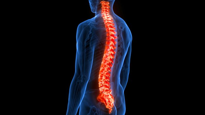
Osteoporosis causes bones to become brittle and prone to fractures. Due to a growing ageing population, the number of patients in Japan is estimated to exceed 15 million in the future. In osteoporosis-related fractures, compression fractures of the spine, known as osteoporotic vertebral fractures, are the most common type of fracture and pose a serious problem, leading to a need for long-term care and a significant decline in quality of life.
One solution is the use of stem cells derived from adipose tissue (ADSCs). These cells are multipotent, meaning they can differentiate into various cell types. Forming ADSCs into three-dimensional spherical clusters, known as spheroids, has been shown to enhance their ability to repair tissue. When these spheroids are pre-differentiated towards bone cells, they can promote bone regeneration.
Bone regeneration in rats with osteoporosis
Bone regeneration is the process by which bone tissue repairs or grows after injury, disease, or degeneration. Naturally, when a bone fractures, the body initiates a series of steps: inflammation, the formation of a soft callus, hardening into new bone, and finally remodelling the tissue over time to restore strength and shape.
The Osaka Metropolitan University-led research team, involving Graduate School of Medicine student Yuta Sawada and Dr Shinji Takahashi, focused on ADSCs for the treatment of osteoporotic vertebral fractures. They developed ADSCs into bone-differentiated spheroids and combined them with β-tricalcium phosphate, a material commonly used for bone reconstruction, to successfully treat rats with spinal fractures.
The team induced osteoporosis in 33 rats (11 per group), creating defects in L4 and L5 vertebrae. Groups included osteogenic spheroids, undifferentiated spheroids, and β-tricalcium phosphate alone. Bone regeneration was assessed using micro-CT, histology, and biomechanical testing at four and eight weeks.
The researchers found that bone regeneration and strength significantly improved in rats transplanted with the complex. Furthermore, they also found that the genes involved in bone formation and regeneration were also activated.
“This study has revealed the potential of bone differentiation spheroids using ADSCs for the development of new treatments for spinal fractures,” Sawada said. “Since the cells are obtained from fat, there is little burden on the body, ensuring patient safety.”.
“This simple and effective method can treat even difficult fractures and may accelerate healing,” Dr Takahashi added. “This technique is expected to become a new treatment that helps extend the healthy life of patients.”


