Cambridge scientists grow ‘hematoids’, embryo-like structures from stem cells that generate blood stem cells after two weeks
Researchers at the University of Cambridge have developed a lab-grown embryo-like model called “hematoids”, which self-organises from stem cells to mimic very early human development. After about two weeks, these hematoids begin producing blood stem cells capable of becoming red blood cells, white blood cells, and immune cells.
The findings are published today in the journal Cell Reports.
Developing lab-grown human stem cells
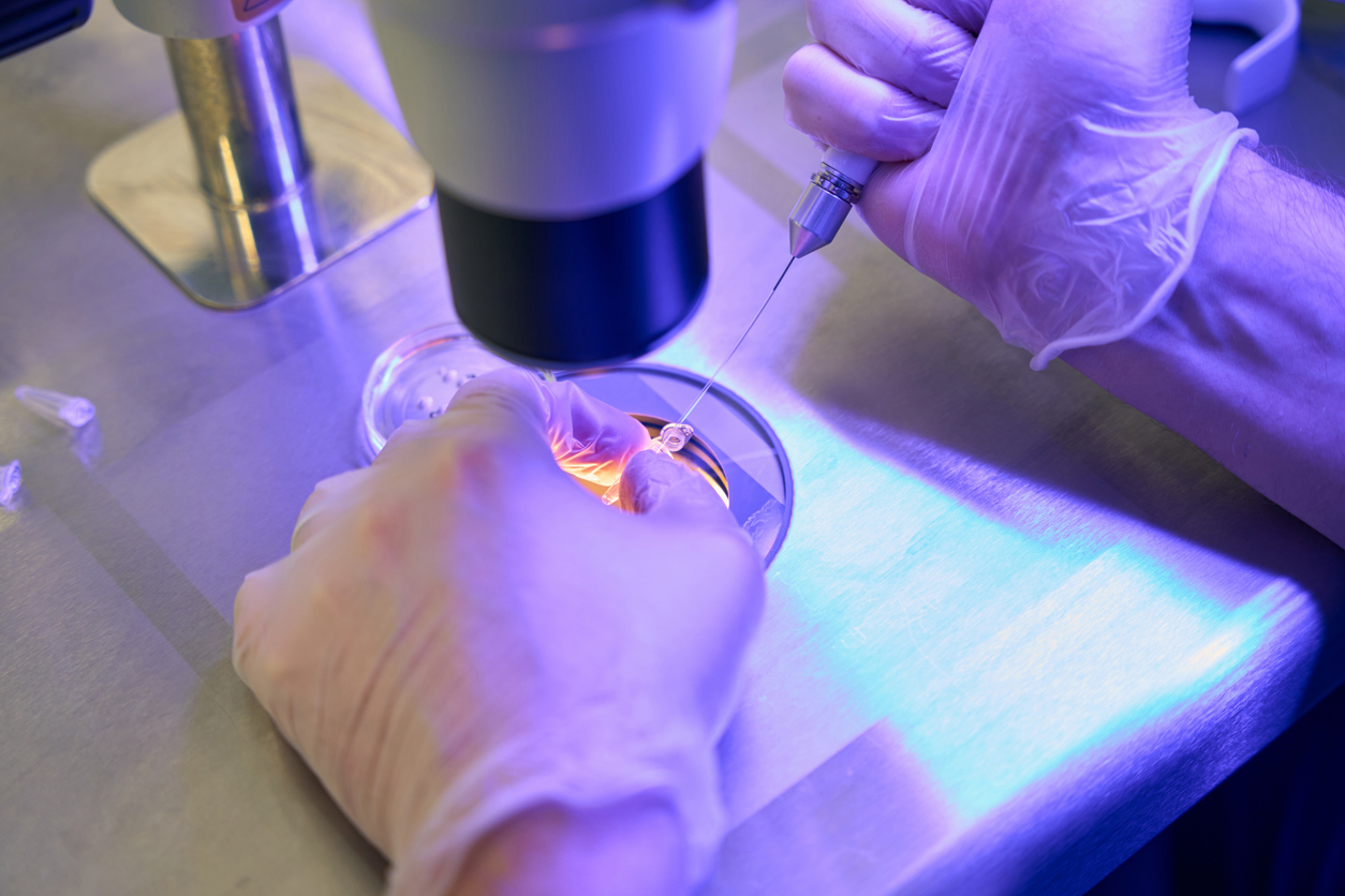
The researchers used human stem cells to create three-dimensional embryo-like structures that replicate certain aspects of very early human development, including the production of blood stem cells.
Human blood stem cells, known as hematopoietic stem cells, are immature cells that can develop into any type of blood cell, including red blood cells that carry oxygen and various kinds of white blood cells. These have high potential for improved understanding of blood formation, unlocking insight into long-lasting blood stem cells for transplants.
The embryo-like structures, named ‘hematoids’ by the scientists, start producing blood after two weeks of development in the lab, acting similarly to human embryos. However, hematoids differ from real human embryos in many ways. They cannot develop into them because they lack several embryonic tissues, including those that support the yolk sac and placenta, which are necessary for further development.
Unlike other lab methods that rely on added growth proteins, this new approach mimics natural development using a self-organising embryo-like model, where cells create their own environment to produce both blood and beating heart cells.
Mimicking human foetal blood development in the lab
The new human embryo-like model simulates the cell changes that occur during the very early stages of human development, when our organs and blood system first begin to form.
The team observed the emergence of the three-dimensional hematoids under a microscope in the lab. By the second day, these self-organised into three germ layers, called the ectoderm, mesoderm, and endoderm, all crucial for shaping every organ and tissue in the body.
By day eight, beating heart cells had formed, which eventually give rise to the heart in a developing human embryo. By day 13, the team observed red patches of blood appearing in the hematoids.
Dr Jitesh Neupane, a researcher at the University of Cambridge’s Gurdon Institute and first author of the study, said: “It was an exciting moment when the blood red colour appeared in the dish – it was visible even to the naked eye.”
He added, “Our new model mimics human foetal blood development in the lab. This sheds light on how blood cells naturally form during human embryogenesis, offering potential medical advances to screen drugs, study early blood and immune development, and model blood disorders like leukaemia.”
Advancing human development knowledge
Stem cell-derived embryo models are vital to advancing knowledge of early human development.
The blood cells in hematoids develop to a stage equivalent to four to five weeks of human embryonic growth, a period impossible to observe in real embryos after implantation. The research followed strict ethical regulations and received full approval, demonstrating the responsible and ethical nature of the scientific community. The team has patented the discovery through Cambridge Enterprise to help translate it into real-world medical and societal benefits.


