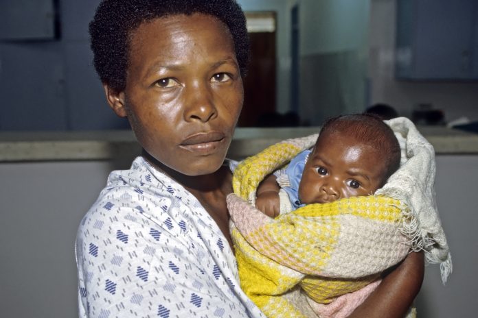
Yuping Wang and David F. Lewis, from Louisiana State University Health Sciences Center, share with us their research on vitamin D and preeclampsia, including a promising option to improve maternal and foetal health
Vitamin D deficiency is a newly recognised risk factor for preeclampsia. Drs Yuping Wang and David F. Lewis at Louisiana State University Health Sciences Center in Shreveport (LSUHSC-Shreveport) are exploring ways of vitamin D to protect placental cells by targeting vitamin D receptor (VDR), a promising option to improve maternal and foetal health.
Preeclampsia, characterised with increased maternal blood pressure and the presence of proteinuria after 20 weeks of gestation, is a serious complication in human pregnancy. This pregnancy disorder is one of the leading causes of maternal and foetal morbidity and mortality worldwide. Although the cause of preeclampsia is not clear, recent studies have shown that low maternal vitamin D levels are associated with a higher incidence of preeclampsia. Therefore, it is considered that vitamin D deficiency is a risk factor for this pregnancy disorder.
Since placental cell dysfunction plays significant roles in preeclampsia development, the research at LSUHSC-Shreveport aims to address the roles of vitamin D in placental cells (trophoblasts, foetal vessel smooth muscle cells (VSMCs) and endothelial cells) and to better understand how vitamin D, a pleiotropic hormone, could ameliorate or rescue placental cell dysfunction in preeclampsia.
Vitamin D metabolic system in placental cells
The key molecules that engage in synthesis and degradation of vitamin D include vitamin D binding protein (VDBP, that carries and delivers 25(OH)D and 1,25(OH)2D3 to target cells and tissues), 25-hydroxylase (converts cholecalciferol to calcidiol, 25(OH)D), 1α-hydroxylase (converts calcidiol to calcitriol, 1,25(OH)2D3), 24-hydroxylase (degradates both 25(OH)D and 1,25(OH)2D3) and vitamin D receptor (VDR). 1,25(OH)2D3 is the bioactive form of vitamin D.
After binding to its receptor VDR and forming heterodimer with other nuclear hormone receptors, the complex then binds to vitamin D response elements (VDRE) in the promoter of genes, in which it regulates and subsequently elicits downstream vitamin D biological actions. Therefore, VDR is an absolute determinant of vitamin D bioactivity.
To study vitamin D metabolism in the human placenta, Drs Wang and Lewis team demonstrated that placental cells are enriched with all the elements that are involved in the synthesis and catabolism of vitamin D.
Their results showed:
1) VDBP and VDR are strongly expressed in placental trophoblasts throughout pregnancy;
2) Vitamin D synthases (25-hydroxylase and 1α-hydroxylase) are not only in trophoblasts and VSMCs but also present in foetal vessel endothelial cells, especially in the third trimester/term placentas, while vitamin D catabolic enzyme (24-hydroxylase) is weakly expressed in placental cells throughout pregnancy and;
3) VDR expression is inducible by 1,25(OH)2D3 in trophoblasts, VSMCs and foetal vessel endothelial cells. These findings indicate that placental cells could produce and store vitamin D to maintain its homeostasis within the placental microenvironment and to provide and transfer prehormone (25(OH)D) from the maternal circulation to the foetus to support the growing foetus’ requirements.
Their works further demonstrated that in preeclamptic placentas, trophoblasts had altered levels of vitamin D synthesis and catabolic enzymes, along with a reduced amount of VDR. Their studies also demonstrated these changes seen in trophoblasts could be induced by mimicking preeclamptic condition induced by oxidative stress in the laboratory.
Could vitamin D improve placental cell function in preeclampsia?
It is known that not having enough vitamin D during pregnancy increases the risk of preeclampsia, but how vitamin D is able to affect placental cell function and whether vitamin D could reverse any of the alterations associated with placental cell dysfunction in preeclampsia are less clear. To answer the questions, the team took the approach to look into the most relevant adverse events that occur in placental trophoblasts in preeclampsia: increased oxidative stress and superoxide generation, increased vasoconstrictor thromboxane and inflammatory cytokine production and increased microparticle release.
Using primary trophoblasts from the human placenta, they demonstrated that bioactive vitamin D, 1,25(OH)2D3 could suppress oxidative stress-induced increase in thromboxane production and superoxide generation, which is due to the ability of vitamin D to inhibit cyclooxygenase-2 (COX-2) upregulation since both thromboxane and superoxide are metabolites of cyclooxygenase pathway. They also demonstrated that 1,25(OH)2D3 could suppress oxidative stress-induced microparticle release by placental trophoblasts via preservation of eNOS expression and inhibition of caspase-3 cleavage and ROCK1 activation in trophoblasts. These findings are important because thromboxane is a potent vasoconstrictor and suppression of thromboxane production could reduce vascular contractility and improve placental blood perfusion.
Moreover, suppression of microparticle release would diminish maternal vascular inflammatory response induced by trophoblasts-derived harmful stimuli. In addition, their data also show that 1,25(OH)2D3 could inhibit inflammatory cytokine TNFα, IL-6 and IL-8 production. Their work, combined with others of suppression of sFlt-1 production by vitamin D, holds a promise that vitamin D has the ability to improve and restore trophoblast function in preeclampsia.
In addition to trophoblasts, they also proved favourable effects of 1,25(OH)2D3 on foetal vessel endothelial cells and found that bioactive vitamin D could promote superoxide dismutase (CuZn-SOD) expression, an essential antioxidant to dismutate superoxide radicals. Their recent study also revealed that 1,25(OH)2D3 could stimulate endothelial cells to produce more miRNA-126, an endothelial specific miRNA that exerts anti-inflammatory activities to govern vascular integrity.
The team also studied vitamin D effects on placental VSMCs and found that 1,25(OH)2D3 could inhibit angiotensin II-induced placental VSMC contraction. Similar to that of losartan, an angiotensin II receptor – 1 (AT-1) blocker, by inducing a signalling mechanism called phosphorylation of the myosin phosphatase target subunit 1 (MYPT1) pathway molecule. This is a key player involved in myosin light chain phosphatase (MLCP) activation that is essential for VSMC relaxation.
Could VDR hold a key to improve trophoblast function in preeclampsia?
The actions of bioactive vitamin D are mediated by VDR, a ligand-activated transcriptional factor that functions to control target gene expression. VDR expression is inducible in placental cells. Therefore, the amount and the activity of VDR present in placenta cells, especially in trophoblasts, are critical to determining the biological effects of 1,25(OH)2D3 derived from the maternal circulation, as well as produced by trophoblasts. However, the amount of VDR on trophoblasts is reduced in preeclampsia. Lack of VDR expression causes trophoblasts unable to respond to 1,25(OH)2D3 properly.
Hence, one of the goals of the research projects of Drs Wang and Lewis research is to find a way to promote VDR production/expression and increase VDR activity in trophoblasts to correct and restore trophoblast function and subsequently, to reduce the incidence or severity of this pregnancy disorder.
David F. Lewis, MD, FACOG, MBA
Professor and Chairman
dlewi1@lsuhsc.edu
Yuping Wang, MD, PhD
Professor
ywang1@lsuhsc.edu
Department of Obstetrics and Gynecology
Louisiana State University Health Sciences Center – Shreveport
Tel: +1 (318) 675 5379
*Please note: This is a commercial profile


The prevalence of preeclampsia is highest in winter and early spring, implicating sunlight deficiency as a possible cause, and low 25(OH)D levels also predict a doubling of the risk of preeclampsia. , Additionally, newborn children of women at risk for pre-eclampsia were twice as likely as other children to be vitamin D-deficient. This is important, because vitamin D-deficient newborns are likely to develop rickets and suffer from convulsions. Pregnant women, obviously, should be sun-seekers, or at least make sure they get other safe UV exposure when the sun is not available.