
Anna Bogdanova, Professor and Head of Red Blood Cell Research Group at the University of Zurich explains how we know how red blood cells look like and if so, what they tell us
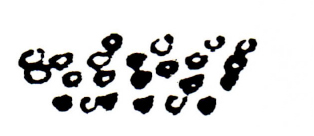
The simplest way to know whether cells exist is to look at them. The first known images of red blood cells date back from 1678, when Jan Swammerdam sketched blood globules he saw using a magnifying glass of Antonie van Leeuwenhoek. These globules were later viewed at somewhat higher magnifications and more details of them were seen. We still cannot use the predictive power hidden in red cell shapes. RELEVANCE consortium members are working to both decode the information red cell shapes offer us and translate it into improved patient diagnoses.
The RELEVANCE project team is contributing to each aspect of this quest, by working with images of living or fixed cells at a variety of magnifications and conditions. Clinical haematologists are used to performing the analysis of red cell shapes in fixed blood smears. Can we automate these tests to make them free from the subjective attitude of a human observer? Can we change from the analysis of static fixed cells to the monitoring of “living” flowing cells exposed to shear, inflammatory factors and other stressors present in our body?
Here are what our early stage researchers (ESRs) tell us about their progress.
Valeria Rizzuto, Josep Carreras Leukaemia Research Institute, Barcelona, Spain (ESR3)
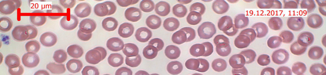
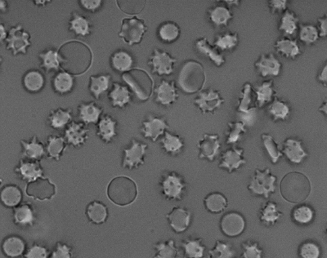
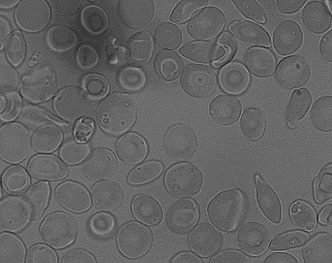
The evaluation of RBC morphology is fundamental since aberrations of RBC shape can provide essential information in establishing a differential diagnosis of anaemia. Here are the challenges we face.
The red cells of patients with rare types of anaemia are intolerant to transportation that have severe impacts on cell morphology. Thus, it would be ideal to perform morphological analysis at the spot, with as little volume as possible and as little technical and human errors and inconsistencies as we can possibly afford. We need more advanced, robust and reliable tests coupled to new approaches in analysis to detect red cell shapes that we plan to develop together with partners from Arivis AG. We currently use peripheral blood smears counting the number of cells with abnormal shapes. Together with our biologist-partners, we plan to get more information about cell shape features in fixed and native red cells, by introducing new parameters to describe cell morphology and by implementing new tests to observe the morphological response to stress.
Greta Simionato, Saarland University, Saarbrücken, Germany (ESR4)
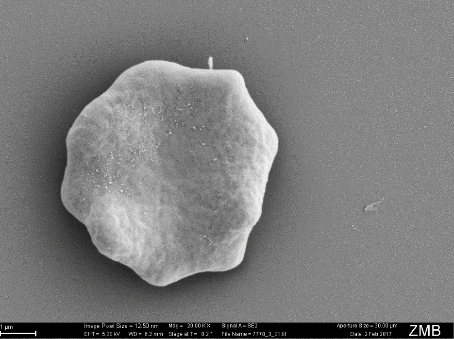
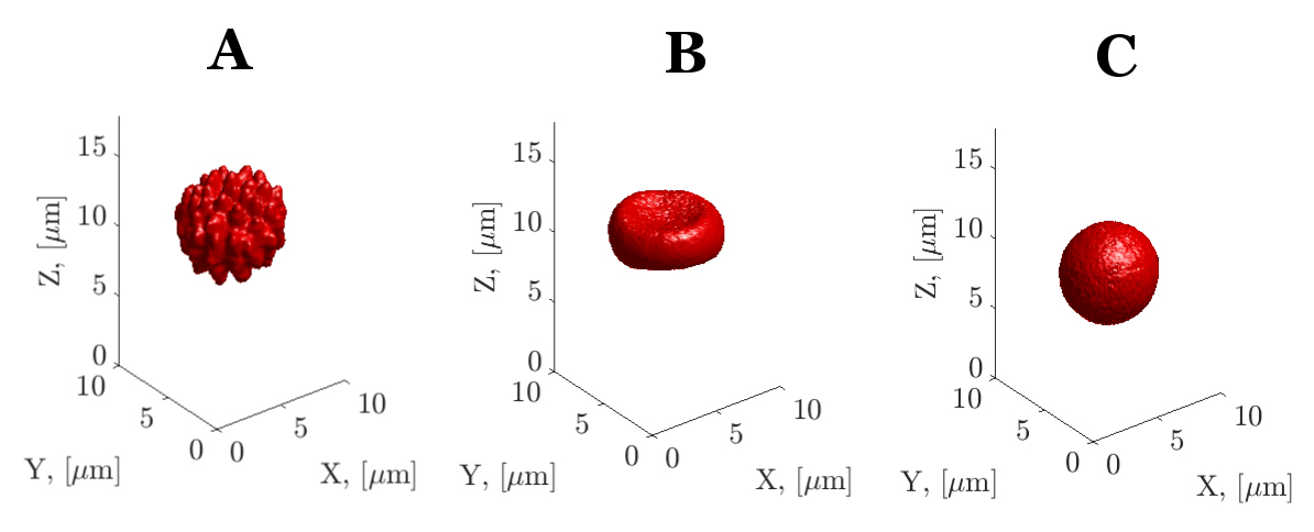
We believe that studying shapes provides a fast and inexpensive approach to learn more about the patients’ condition and molecular causes of disease. We are currently working on the improvement of imaging techniques. We apply confocal microscopy for transferring 2D- into 3D-images of cells. We develop new approaches for image analysis that include the extraction of quantitative parameters describing distinct elements of cell shape such as roundness, border regularity, volume and surface area. We explore the predictive power of these parameters for diagnosis and severity prognosis for patients with rare types of anaemia. We develop automatic cell shape recognition and a classification system, using a neural network approach to achieve fast, unbiased and more sensitive cell shape analysis.
Sinja Novosel, University of Zurich, Zurich, Switzerland (ESR7)
Our role within the consortium is to bridge the research to the clinical and industrial demands. We develop a set of tests to challenge red blood cells and assess their responses to physiological stressors, such as acidic pH, Ca2+ uptake, or shear stress. These tests will serve better visualisation of the abnormalities and red cell membrane architecture that cannot be clearly monitored in cells “at rest”. We decipher the links between the mutations in proteins building membrane skeleton and the specific shapes cells prefer to acquire.
We also study the biological mechanisms behind heterogeneity in red cell shapes and properties. These tests will be then implemented in a new prototype of a microfluidic device that will be used for automated cell shape analysis. This device will address hydration state, deformability and membrane stability of individual red blood cells. Information on the physiological and pathological “meaning” of red cell shape preferences and heterogeneity of cell shapes will be then implemented for the development of the algorithms for red cell shape analysis and its interpretation for clinical applications.
Niamh Kilcawley, Epigem Ltd, Redcar, UK (ESR1)
A major challenge that we are faced with is the volume limitations associated with the screening of blood from newborn infants and neonates. For this, it is necessary to only use microliter sized samples. The first two working prototypes of a microfluidic instrument has been developed with the aim to run these low volume blood samples for the screening and detection of rare anaemia and are currently on probation in a clinical lab in Division of Pediatric Hematology of Emek Medical Center in Afula, Israel and with the team of Red Blood Cell Research Group at the University of Zurich. For me, this is a new phase in my work – a transition from building to testing. Next step will involve improving the microfluidic system. Together with the other partners, we will evaluate the predictive potential of the tests.
Niamh (on behalf of other ESRs)
We believe that the RELEVANCE project will make a massive difference to so many people. By studying red cells in depth, we can progress not only our knowledge of red cells and their mutations, but also progress the treatments and procedures for improving the quality of life for these patients. The interaction between all the sciences, universities, hospitals/clinics and SMEs gives all the ESRs access to such a broad range of education – that is not typical in a normal PhD situation. It also gives us connections to each other and the possibility of great collaborations which is very good ‘life learning’ for anyone who wants to pursue a career in science.
Please note: this is a commercial profile
Anna Bogdanova
Professor and Head of Red Blood Cell Research Group
University of Zurich
Tel: +41 (0)44 635 8811


