
Experts from the European Laboratory for Non-Linear Spectroscopy and the Neuroscience Institute reflect on cerebral blood flow (CBF)
The circulatory system of our body can be imaginatively described as a highway, through which oxygen and nutrients are carried to feed our organs. About 60,000 miles of blood vessels and a high degree of complexity, flexibility and efficiency make our body’s transportation plan a super engineering work, with no rival among man-made roads. The blood flow ensures all the body districts to be continually supplied with all the substances needed for their activities.
However, loss of blood flow (ischemia) can occur, leading to organ dysfunctions. The severity of these events, depends on the site, with some organs being more sensitive than others to reduced blood flow. In particular, the brain is more depended than other organs on a continuous supply of blood. This dependence is due to the lack of fuel reserves and the high brain’s energy demand. If cerebral blood flow (CBF) is interrupted, brain function ceases within seconds leading to irreversible damage to its cellular constituents within minutes.
Such interruption of CBF can come in different sizes and shapes, with consequences depending very much on the part of the brain affected. In what is called a transient ischemic attack (TIA), the blood supply to a part of the brain fails temporarily. The symptoms of TIA resolve within 24 hours and include sudden dimming or loss of vision, aphasia, slurred speech and mental confusion.
A prolonged cut off of blood supply is called stroke and can lead to irreversible damage. The most common symptom of a stroke is sudden weakness or numbness of the face, arm or leg, most often on one side of the body. Other symptoms include confusion, difficulty speaking or understanding speech, difficulty seeing with one or both eyes, difficulty walking, dizziness, loss of balance or coordination, severe headache, fainting or unconsciousness.
Moreover, a very severe stroke can cause sudden death. The World Health Organization estimated in 2012 cerebrovascular accident (another name for stroke), as the second leading cause of death and the third leading cause of disability worldwide.
Nowadays imaging techniques such as magnetic resonance angiography (MRA) and computer tomography angiography (CTA) represent important diagnostic means to identify vascular abnormalities or evaluate vessels occlusions. MRA and CTA yield images of brain vasculature with minimal invasiveness. For this reason, they keep a relevant role in the diagnosis of vascular diseases like stroke.
In addition to diagnostic use in humans, these methodologies have been applied in pre-clinical settings to obtain full datasets of whole brain vasculature. Time of flight (TOF) angiography, for instance, is a non-invasive MR method which allows the visualisation of blood flow in whole mouse brains with a resolution close to 1 mm. Contrast-enhanced MRA and microscopic computed tomography (µCT) take advantage of contrast agent injected into the blood flow to visualise smaller vessels (10 times smaller than TOF-MRA) in whole rodent brain.
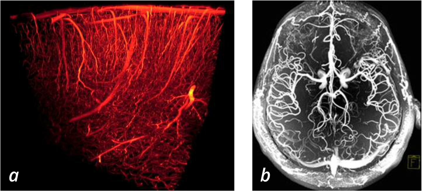
On the other hand, these approaches are profoundly wanting on a microscopic level, which means that only large vessels are detectable (Fig.1a). The resolution needed for visualising single capillaries is instead achievable with optical microscopy (Fig.1b). Thanks to the advances achieved in optical technologies in the last years, we are now starting to unveil the intricate organisation of the brain vascular network on a micrometric scale.
Imaging techniques
Optical imaging techniques like two-photon fluorescence or light-sheet microscopy allow evaluation of changes in the vascular organisation, at the level of the capillary network in animal models of vascular impairments. In combination with recently developed optical clearing techniques, these methods provide fine morphological vascular details in large volumes of tissue, thus coupling high-resolution with brain-wide analysis.
This level of analysis is of remarkable importance to improve our understanding of metabolic processes, underlying both physiological and pathological conditions. Nevertheless, such a fine analysis requires considerable efforts to be accomplished in large volume. Indeed, with the purpose of obtaining a complete understanding of the brain vascular network, methodological developments involving computational analysis and microscopy technologies are currently ongoing.
The specific features of optical microscopy make it also a complementary tool when associated with other imaging methods. For instance, the coupling between MRA imaging with the latest optical methodologies will allow us to associate functional studies with a detailed vascular structure. Ultimately, the approaches described above will permit to step forward towards the achievement of whole-brain vasculature network reconstruction.
Such achievements will have implications – ranging from the generation of better diagnostic tools to the availability of innovative procedures to be used in the field of brain research – eventually leading to new treatments of vascular-related pathologies including cancer, stroke, and Alzheimer’s disease.
The tremendous efforts against this kind of pathologies are justified by the strong impact on the society. For instance, stroke and cancer often affect individuals at the peak of their productive life, while the spreading of neurodegenerative disorders entails an increasing burden on public health. It emerges that beyond the personal and familiar difficulties, these conditions have an enormous effect on countries’ socio-economic development. For such reasons, improving our knowledge about physiological brain activities and abnormalities is necessary, with the purpose of getting effective therapies to up to now incurable brain related dysfunctions. In this frame, the use of optical techniques can be a valid strategy to extract fine morphological information and to better define disease phenotypes. This will potentially lead to the identification of new brain alterations associated with specific pathologies to be used as new targets for therapeutic treatments.
Please note: this is a commercial profile
Antonino Paolo Di Giovanna
Researcher
European Laboratory for
Non-Linear Spectroscopy
Tel: +39 (0)5545 72267
Anna Letizia Allegra Mascaro
Researcher
Neuroscience Institute,
National Research Council
Tel: +39 (0)5545 72504
Francesco Saverio Pavone
Full professor
European Laboratory for Non-Linear
Spectroscopy and Department of Physics
University of Florence
Tel: +39 (0)5545 72480


