
Dr. James E Goldman and Dr. Osama Al-Dalahmah from the Department of Pathology and Cell Biology at Columbia University, discuss the importance of brain banking from individuals with Huntington’s disease
In an article in Open Access Government in 2021, we described a new experimental strategy to study the brain tissues that have graciously been donated for research by Huntington’s disease (HD) patients and their families. We discussed a technique called single nuclei RNA sequencing (snRNASeq), in which we isolate the nuclei from thousands of cells from a piece of frozen brain tissue and extract the RNA from these nuclei to determine what genes are being expressed in individual cells of the tissue.
By comparing our results from HD brains to those of individuals without neurologic or psychiatric disorders (control donors) we have found many differences in gene expression due to the presence of the mutant protein causing HD. We have found changes in all of the different cell types that make up the human brain, neurons (of different types), astrocytes (cells that control the levels of ions and neurotransmitters and that help neurons to survive), oligodendrocytes (the cells that make myelin, the insulating substance of axons), and microglia (the immune cells of the brain).
Understanding the cellular and molecular mechanisms of neurological disorders requires access to human brain tissues
Some of these changes in gene expression are positive, reacting to the alterations caused by the toxic mutant protein and helping cells to survive these alterations. Some of the changes are deleterious, provoking reactions that further damage the tissue and prevent normal functioning.
To accomplish this research, we must have access to brains from well-characterized donors with or without Huntington’s disease, which were obtained at autopsies and have been processed meticulously for basic investigations. We cannot isolate nuclei from tissues that have been preserved in a fixative. Thus, we must rely on brains that had been harvested as soon as possible after death, then carefully dissected in the fresh state to generate blocks from specific areas. These blocks are immediately frozen and kept at a very low temperature to prevent degradation.
The importance of brain banks and availability for research
We have been extremely fortunate to work with the New York Brain Bank (NYBB) at the Columbia Irving Medical Center, where we conduct our studies. The NYBB was established in our Neuropathology Division and with its support in 2001 under the guidance of Dr. Jean Paul Vonsattel, whose focus of interest is HD and other neurodegenerative disorders. The new Director of the NYBB is Dr. Andrew Teich.
When a fresh or an unfixed brain is made available for research, we divide the brain in half, down the middle. From one half we dissect 50 or more precisely selected blocks, each about one square inch, and each including specific areas of interest. The blocks are placed between Teflon-coated plates), frozen in liquid nitrogen vapor and then stored in a freezer at -80o C. In addition, we collect many other pieces that are crushed in liquid nitrogen, and stored in 2 ml vials; these samples are suitable for research that do not require integrity of the cellular morphology.
Each fresh frozen block and each crushed frozen parenchyma containing vial receives a barcode, unique to that individual brain. Relevant information is entered into a computer database, including the barcode, and the freezers, shelves and boxes where the samples are stored. This allows us to access critical information easily. The other half of the brain is immersed in a formalin solution, which is a standard fixative. A subset of the same areas as those obtained from the unfixed half of the brain are collected from the fixed half brain and placed in cassettes for embedding in wax.
We then cut and stain thin sections from the fixed pieces and examine them under the microscope to render a detailed neuropathological diagnosis. The New York Brain Bank provides optimally prepared and diagnostically categorized human brain samples to neuroscientists worldwide. The Bank mails many hundreds of samples each year.
RNA sequencing and single cell nuclei
For Dr. Dalahmah’s and my work, we require several qualities from the banked tissues. First, for RNA sequencing, the RNA of the tissue must be well preserved. RNA will break down after a patient’s death, after which RNA sequencing will not be possible. The frozen specimens in the Bank contain very well-preserved RNA.
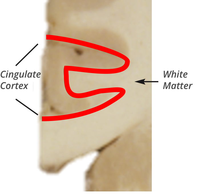
Second, we are interested in examining single cell nuclei in specific regions of the brain. In the case of Huntington’s Disease we have isolated nuclei from the cingulate cortex, the caudate nucleus, and the nucleus accumbens, and are planning to examine further regions. To do this we must first isolate the specific area we wish to analyze. The brain is a complex structure and each of the specimens may contain more than the specific region from which we want to isolate nuclei. For example, the specimen of the cingulate cortex contains both cortex and the subcortical white matter (the area under the cortex that contains the axons of cortical neurons). The anatomy of the pieces is clear in the frozen specimens, so that we are able to dissect out only those regions we want to analyze (Figure).
Third, we must be able to cut very thin slices from the frozen tissues to perform a variety of special stains. It is important in these sections to be able to see the microscopic anatomy and the individual cells clearly and without any distortion. The method the Bank uses to freeze the tissues preserves the structure of the tissue very well.
We have been fortunate to have access to brains stored at the NYBB. Indeed, neuropathologists from other medical centers have visited our Brain Bank to learn the protocols to help them establish their own repositories or upgrade current ones. We would strongly urge governments and institutions that want to study neurological diseases using these sophisticated molecular techniques to establish high quality brain banks or to support those that are already in place.
The Figure shows a piece of brain tissue that includes the cingulate cortex and underlying white matter. We can dissect out the cortex free of other areas along the red line so that the sample of cortex is not contaminated by white matter.
Please note: This is a commercial profile
© 2019. This work is licensed under CC-BY-NC-ND.


