The role of advanced technologies in healthcare, including the work of the National Institute of Biomedical Imaging and Bioengineering (NIBIB) in this area, is placed under the spotlight by Open Access Government
On the website of, National Institutes of Health (NIH), we learn that for most of the history of medicine, doctors have relied on their senses – that is vision, hearing and touch – to diagnose illness and monitor a patient’s condition.
However, NIH’s investment in research over the period of a whole century has helped to completely change medical diagnostics. The new technologies of today allow doctors to find out an increasing amount of detailed information about both the progression and treatment of disease and can even offer personalised treatment based on a patient’s genes, we discover.
In addition, NIH-funded scientists have helped to pioneer the development of magnetic resonance imaging (MRI), but they have also made important strides toward uncovering the medical potential of stem cells and as such, they have developed new tools for genome sequencing. With these advanced technologies in place, scientists are finding out how genes affect human health and how a genetic approach can help doctors tailor treatments and prevention strategies for each patient. Certainly, millions of individuals have already been touched by the exciting era of personalised medicine that has grown directly from this research, we learn. (1)
Advances in medical imaging
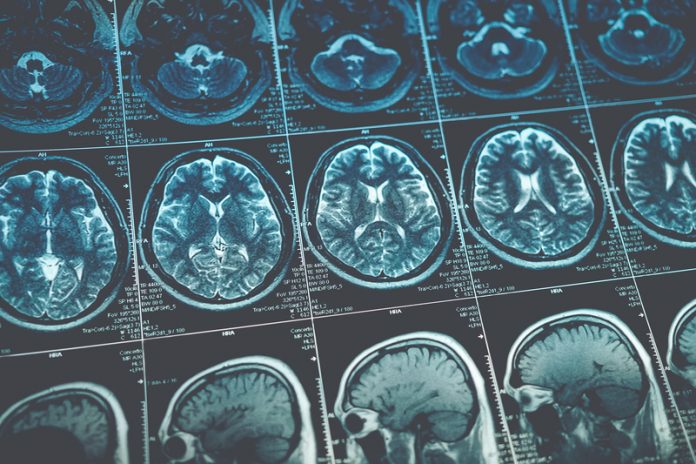
One area that concerns the NIH is medical imaging, an area they are thoroughly committed to. Indeed, they have an entire institute, the National Institute of Biomedical Imaging and Bioengineering (NIBIB), who seek to improve health by leading the development, acceleration and application of biomedical technologies.
NIBIB is devoted to integrating the physical and engineering sciences, with the life sciences, to further basic research and medical care. This ambitious aim is achieved through:
- Research and development of new biomedical imaging and bioengineering techniques and devices to improve the prevention, detection and treatment of disease;
- Furthering present imaging and bioengineering modalities;
- Supporting relevant research in the physical and mathematical sciences;
- Encouraging research and development in multidisciplinary areas;
- Supporting studies to assess the effectiveness and outcomes of new biologics, processes, materials, devices and procedures;
- Developing technologies for the early detection and assessment of health status where diseases are concerned and;
- Developing advanced imaging and engineering techniques for carrying out biomedical research at multiple scales. (2)
Below, we explore just two examples of the excellent research NIBIB is supporting, where medical technologies are concerned.
Thermo-chemotherapy combo eradicates primary and metastatic tumours in mice
In recent news, we find out that bioengineers at NIBIB developed a smart anti-cancer nanoparticle with precisely targeted tumour-killing activity, which was found to be superior to the previously available technologies.
We are told that state-of-the-art nanoparticle features a very sturdy shell, which is capable of carrying large loads of chemotherapeutic drugs through the circulatory system to a tumour – that is without the leakage that can damage healthy tissue. The nanomedicine is photothermal-responsive, so the cancer-killing load of the particle is released only when it enters the tumour cells and is activated by laser light, we discover.
Reported in the February issue of Nature Communications, this cancer-killing technology is the latest and most effective created by members of the Laboratory of Molecular Imaging and Nanomedicine (LOMIN) at NIBIB, which is led by Xiaoyuan (Shawn) Chen, Senior Investigator.
Fluorescent nanoparticles track cancer metastasis to multiple organs
Researchers funded by the NIBIB have developed fluorescent nanoparticles that light up to track the progress of breast cancer metastasis, we find out. They are currently testing the particles in mice, with the hope of one day using them in humans, we are told.
Prabhas Moghe, Ph.D., professor of Biomedical Engineering and Chemical and Biochemical Engineering at Rutgers University and his team are developing nanoparticles that can help identify and track cancer metastasis early on, even when a tumour is really small.
“There are still significant hurdles to be overcome before this imaging technique could be used in humans, but it is an important step in moving optical imaging cancer diagnosis forward,” says Behrouz Shabestari, Ph.D., director of the NIBIB Program in Optical Imaging and Spectroscopy. “It has the potential to help doctors to identify tiny cancer cells and track their spread more effectively, leading to early detection and potentially improved treatment planning.”
As well as tracking the spread of cancer, the nanoparticles could potentially be used to differentiate cancer tissue from healthy tissue for surgeons who are removing tumours. Moghe and his team are hopeful that the technology could be positioned for use in humans within the next 10 years. (3)
Final thoughts
You can find many more examples of how NIBIB is improving human health, by leading the development and accelerating the application of biomedical technologies in the US today, which this article provides a flavour of.
For more information, please visit www.nih.gov.
References
2 https://www.nih.gov/research-training/advances-medical-imaging
Open Access Government


