
Non-Human primates (NHPs) continue to be fundamentally necessary to advance our understanding of the human brain, from its molecular and cellular make up all the way to its systems architecture and how all these mesmerizing components give rise to behavior. This necessity is not changing. It is the curiosity and effort of the human brain to understand itself
To do so, for many studies it uses surrogate brains from other species. One of this species is the rhesus macaque, one of the most studied animal models in the biomedical field. In the foreseeable future this is also not going to change. Many experiments are not possible or allowed in human nor in chimpanzee, its closest relative; hence for obvious reasons, from the ethical to the legal to the plain common-sense similarities between the species, rhesus macaques will continue to be an animal model in high demand.
The 3Rs principles and the MacBrain Resource Center (MBRC) mission
Since the 3Rs proposal, about 65 years ago, to “reduce, replace, and refine” animal use in the framework of the performance of more humane animal studies (Russell and Burch, 1959), a tremendous effort has been devoted to this goal with rather limited success when it comes to the complexities of studying the brain. Complete replacement of animals may not yet be possible but a reduction based on better management of animals already sacrificed or to be sacrificed is within reach. For unviability of complete replacement take for instance electrophysiological studies.
The electrical signal of cells comes from the unequal distribution of ions across the cellular membrane; cells are therefore like batteries and for them to have an energy signature, they must be alive. Dead cells are alike discharged batteries. Therefore, electrophysiological recordings can only be done in live animals, or brain slices with still live cells, that can only be obtained by special protocols; usually, immediately following animal sacrifice. Removal of organ pieces, as sometimes done because of exploratory biopsies or as treatment (e.g., intractable epilepsy) while many times rather inconsequential in many organs are usually a last resource for the brain, with most of the time undesirable sequala, not to mention that these are limited to unhealthy tissue which therefore hinders the study of normal natural electrophysiological behavior.
Organoids and cell cultures are tremendously useful and have produced insightful data, but, have many limitations of which the lack of complex sensory perception and of a consequent behavior are commonly cited.
Animal reduction is however possible in the case of histological studies. But this requires effort, sharing, trust, good communication and a conscious change in behavior. First needed change is the customary practice of using what is required for the immediate research needs while discarding the rest. The practical limitations of preserving tissue can be surmounted with additional resources, e.g., an additional freezer for immediate tissue storage, the possibility of depositing materials into institutional or government run repositories, but, above all, more trust and better communication between researchers.
Some of this is already being implemented, a few government agencies, many institutions, some with and some without governmental support, have created and are managing tissue/organ repositories for the benefit of all. These efforts are to be promoted so that further support ensures their long-lasting benefits. This applies not only to the brain, but to the whole body. Lack of sharing may hence just be a lack of better communication which is not excusable at a time when, in an instant, many people, far away from each other, via social media share what they just had for breakfast with elaborate rhetoric and beautiful images.
Hence, via properly managed communication networks the same can be done for or by scientists using animals in research. Increased communication will increase, trust and sharing.
So, let me share with you that the materials available in the MBRC, in the Department of Neuroscience at Yale University School of Medicine, may, in many instances, constitute a true and very viable alternative to help NHP brain researchers reduce, replace, and/or refine studies that require NHP brain histology. These materials are organized into different Collections (see: https://medicine.yale.edu/neuroscience/macbrain/). Some of it is frozen brain tissue blocks (Collection 7), some are blocks prepared for electron microscopy (Collection 5) and a lot of it is digitized images of tissue already processed (e.g., Collections 1 and 6). Additional details on the tissue Collections, their history, and the effort to make these materials available have been previously published (Duque and Selemon, 2018; Selemon and Duque, 2019; Duque 2023).
It is in this context that the 3Rs principles are at the heart of MBRC mission, it strives to provide a cost-effective means for researchers to conduct de novo studies on NHP brain using materials already in existence and therefore, minimizing or avoiding altogether the need to sacrifice additional animals.
Of course, the extended domain of the mission includes the constant incorporation of new materials into the different collections with the idea that the larger the bank of processed and unprocessed specimens the more useful the resource becomes.
The ART of doing NHP neuroscience research at a fraction of its COST
The following discussion will be developed around the concept of ART COST, a reference to the argument that both science and art are primarily a journey of exploration and that cost is always associated with any enterprise.
Here, ART stands for Availability, Reproducibility, and Transparency. The MBRC certainly fits these attributes. It currently houses seven Collections, all of them publicly Available, to foster Reproducible and Transparent NHP brain research. The Collections are all highly dynamic, with,
for instance, brain tissue blocks left as a biproduct of experiments being added, or used as needed, and newly processed brain sections constantly being digitized and made publicly available.
For scientific advancement to occur, collaborations are usually indispensable; these introduce new ways, new voices, new interpretations, and many times benefit from the use of new technologies; all combined in an environment of complementarity and cooperation, that potentiates different strengths and can bring together different areas of expertise around common or even diverse goals. It is an extraordinary advantage that virtual materials are reusable ad saecula saeculorum without exhaustion.
Hence, any researcher may use the same material at any time, multiple times. The fact that digitized already processed brain sections are viewable and downloadable via the world wide web allows collaborations not possible otherwise. Researchers in different institutions, different countries and different continents can all access the same “raw” virtual materials and together work as equals, without inherent advantages or disadvantages. This level of materials’ Availability to foster friendly scientific cooperation or even competition, is completely Transparent and promotes Reproducibility of results.
Public awareness is part of the Transparency. By removing as much as possible the veil covering the mysteries of what is being done by scientists the public is educated and in a much better position to understand the importance of brain research, and be aware of the responsible, ethical, and very serious commitment of most scientists to use the least number of animals possible and to conduct animal research with the highest standards of care to avoid, as much as possible, any pain, suffering and unnecessary experimental repetition. To increase public awareness, when it is mostly the publics’ money that is being used to conduct animal research, seems responsible, convenient, and due.
Here, COST is used as the acronym for actual Cost, plus Opportunity, Sacrifice, and Translational value. The fact that the MBRC makes the same virtual materials available to all researchers does indeed reduce the financial burden for individual laboratories to access NHP specimens and the Cost associated with processing their large brains. Making the materials available, either expendable materials or everlasting scanned images, reduces the Cost and the need to Sacrifice animals while increasing the Opportunity for any interested researcher to expand or complement what they can already do. Interspecies comparisons are just one example of the multiple ways in which research can benefit. MBRC materials have already afforded, and are likely to continue to afford, brain histological comparisons between different strains of mice and rhesus macaque (e.g., Ding, 2022; Wildenberg et al., 2023). Of course, there are many limitations and one of these limitations may be that only control-untreated animals can be compared.
Nevertheless, timely comparison may shine light on what the case is in primates and help in multiple ways guide further experimentation in mice, or the planning of additional experiments in NHPs. These efforts have tremendous potential to, from very early on, increase the Translational value of all animal experimentation.
Combining the old and the new
What the MBRC has that is old is archived materials that are curated, or can be curated, and perfectly good to continue being explored using new technologies and/ or in combination with newer materials, as illustrated in figure 1. Old stones are still used to build new cathedrals. The complementarity of the information that can be acquired is unique and powerful.
Artificial intelligence can now be used to help analyze materials that were produced decades ago and have been properly preserved. Depending on the study there is either no need or minimal need to sacrifice additional animals. For instance, without a single new animal being sacrificed, new studies on the subplate, the amygdala, the thalamus, and the stria terminalis have been published (Duque et al., 2016; MacDonald and Duque, 2022; Spadory et al., 2022; Sakharkar et al., 2022). In additional new studies, still significantly reducing the number of animals sacrificed, the old materials have been a guide and/ or a complement to research on gyrification, the development of the claustrum, and processes in the prefrontal cortex and areas related to memory and Alzheimer’s disease (Rash et al., 2019, 2023; Li et al, 2023; Yang et al., 2022; Leslie et al., 2021; Galvin
et al., 2021; Datta et al., 2020, 2021).
Learning the hard way
The COVID-19 pandemic helped governmental agencies and the public in general realize that current NHP experimentation is essential to assure human survival. Development and testing of appropriate vaccines in NHPs were fundamental for validation before they could be used on large numbers of humans worldwide in what can be considered an existential threat of global proportions. The necessary confinement also helped speed up the technologies of virtual communication including those that can be used for research and therefore very clearly indicated the tremendous value of virtual access to archived research materials.
Let us not forget that NHPs have been key in the development of other vaccines (polio, measles, Ebola) and more recently in the development of deep brain stimulation for the treatment of Parkinson’s’ disease. Therefore, better management of what is obtained by each sacrifice can potentially decrease the need for future sacrifices.
Concluding Remarks
At this time, a time of extraordinary advances in computer science, robotics, artificial intelligence, and general technical capabilities, the point has been reached when new and emergent technologies can be used to explore large data banks in a faster and more efficient way. Increased data storage capability and data acquisition and transmission speed, more than ever before justify and facilitate the use of archived materials to decrease the number of animals needed to be sacrificed for biomedical research and give investigators additional tools to advance human and animal health-related research.
These tools are particularly useful in the case of NHPs which are animal models harder
to secure and more expensive to sustain. Widespread communication helps the dialogue between scientists, governmental agencies, legislators, and public in general for the advancement of better, clearer, properly regulated and more transparent research agendas that do not antagonize the scientists and their work and end up hurting the very animals we have a duty to protect. For further general information see also Homberg et al., 2021; Nonhuman Primate Models in Biomedical Research: State of the Science and Future Needs, 2023.
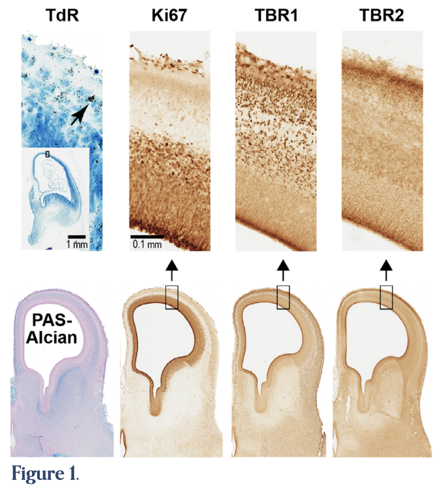
Figure 1. (TdR) A brain section from a tritiated thymidine (TdR) case (011173) done in Pasko Rakic’s laboratory 50 years ago in which a pregnant monkey was injected with TdR at gestational day 40 and the brain of the embryo, obtained by C-section 7 days later (E47, Collection 1 of the MBRC), was processed by autoradiography to follow the fate of the labeled cells. The arrow points to a heavy deposit of silver grains on the nucleus of a cell whose precursor was in the S-phase of the cell cycle, i.e., at the time of injection (i.e., the cell was born at the time of injection). (PAS-Alcian, Ki67, TBR1, TBR2), are different types of markers recently done by histo- and immunohistochemistry on brain tissue from an E49 embryo (Rakic lab).
Periodic Acid-Schiff Alcian blue stain (PAS-Alcian) is used for the visualization of the extracellular matrix, particularly abundant in the subplate. Brain Specific T-box transcription factors 1 and 2 (TBR1, TBR2), are neuron-specific transcription factors whose derailment during development is associated with different brain disorders and later with the consequent intellectual disabilities. They are commonly used to label what will be projection neurons and intermediate progenitors. Ki67 is primarily a proliferation marker.
Funding
Funding for the MBRC is provided by the National Institute of Mental Health (grant RO1MH113257 to Dr. Alvaro Duque).