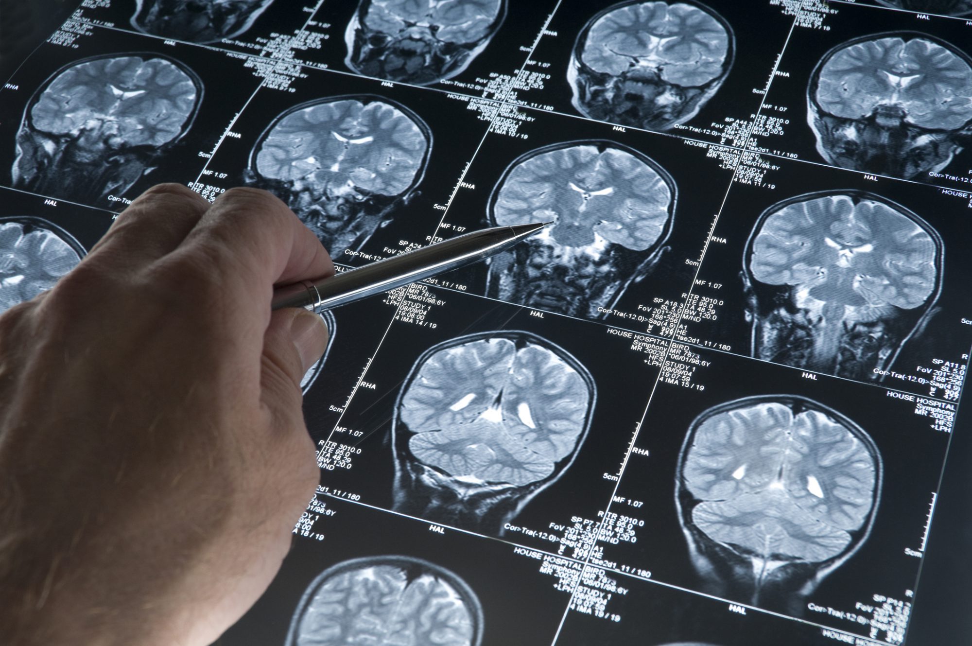
Researchers have introduced a non-invasive method for determining the malignancy grade of glioma tumours
This discovery was led by Professor Yasuchika Hasegawa and Professor Shinya Tanaka of the Institute for Chemical Reaction Design and Discovery (WPI-ICReDD) at Hokkaido University and offers hope to brain tumour patients.
Cancer-grade probing system
The cancer-grade probing system (GPS) uses a water-soluble, luminescent europium complex, creating a non-invasive alternative to current invasive and risky methods of assessing brain tumour malignancy.
Gliomas, comprising 26.3% of all brain cancers, present a significant challenge in determining malignancy grades crucial for selecting the most effective cancer therapy. The team’s approach involves introducing the water-soluble europium complex to model cells replicating glioma, allowing for a nuanced evaluation of malignancy grades.
The key to their success is measuring changes in the complex’s characteristic red-light emission lifetime.
Conducting the study
The study involved testing three different model cells simulating distinct grades of malignancy. Within the initial three hours of introducing the europium complex, the researchers observed more pronounced changes in the lifetime of light emission in cells with higher malignancy grades.
This breakthrough could pave the way for non-invasive tests to determine patient tumour malignancy, eliminating the need for invasive procedures with high risks of complications.
Professor Hasegawa highlighted the originality of their approach, stating, “Visualisation of cancer cells using luminescent complexes has previously been reported, but we hypothesised that the photophysical signals sent by such complexes in cancer cells might reflect internal information from the cancer cells.”
Creating water-soluble amino acids
The researchers first modified its properties to make the europium complex water-soluble and stable among amino acids in the cell culture medium.
When added to the cell culture medium, the complex initially forms aggregates with itself. These aggregates break into single molecules upon interaction with model tumour cells, rapidly entering the cells and inducing structural changes that affect the complex’s red-light emission lifetime.
“Brain tumours occur in 4.6 out of every 100,000 people in Japan”
The differences in emission lifetimes were linked to varying tumour activity and growth processes among different malignancy grades. This suggests that the method could allow continuous detection of tumour activity, providing crucial information for doctors when deciding on appropriate treatment strategies.
Professor Tanaka emphasised the potential impact on patients, saying, “Brain tumours occur in 4.6 out of every 100,000 people in Japan, and the 5-year survival rate is 16% for the most malignant grade 4 type of glioblastoma, which is an aggressive type of glioma brain tumour. The malignancy evaluation method we developed may be able to benefit these patients in the future.”
Editor's Recommended Articles
-
 Must Read >> AI unlocks cancer treatment secrets
 -
-